Download
1 / 8
80 likes | 104 Vues
In the present study, the wound-healing activity of some substituted hydroxytriazenes was investigated. Excision, resutured incision and dead space wound models were used to evaluate the wound-healing activity of substituted hydroxytriazenes. In excision wound model, treatment was continued till the complete healing of the wound, in incision and dead space wound models the treatment was continued for 10 days. For topical application, 5% w/w ointment of hydroxytriazenes was prepared in 2% sodium alginate and for oral administration hydroxytriazenes dissolved in DMSO, at dose 5 mg/kg were used. In excision and incision wound models, the control group of animals was left untreated and in dead space wound models the animals were treated with DMSO (1ml/kg). The healing of the wound was assessed by the rate of wound contraction, period of epithelialisation, skin breaking strength, granulation strength, dry granulation tissue weight and hydroxyproline estimation. Except the parent compound (HD-1), rest of the hydroxytriazenes (HD-2 and HD-3) significantly promoted the wound-healing activity in all the three wound models. Substituted hydroxytriazenes (HD-2 and HD-3) promote the wound-healing activity, whereas the parent hydroxytriazene compound (HD1) does not do so.

E N D
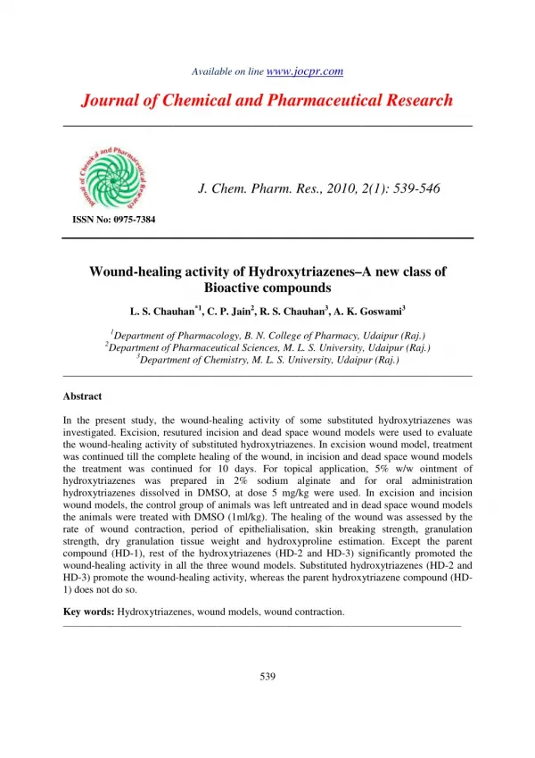
Available on line www.jocpr.com Journal of Chemical and Pharmaceutical Research ____________________________________________________ J. Chem. Pharm. Res., 2010, 2(1): 539-546 ISSN No: 0975-7384 Wound-healing activity of Hydroxytriazenes–A new class of Bioactive compounds L. S. Chauhan*1, C. P. Jain2, R. S. Chauhan3, A. K. Goswami3 1Department of Pharmacology, B. N. College of Pharmacy, Udaipur (Raj.) 2Department of Pharmaceutical Sciences, M. L. S. University, Udaipur (Raj.) 3Department of Chemistry, M. L. S. University, Udaipur (Raj.) ______________________________________________________________________________ Abstract In the present study, the wound-healing activity of some substituted hydroxytriazenes was investigated. Excision, resutured incision and dead space wound models were used to evaluate the wound-healing activity of substituted hydroxytriazenes. In excision wound model, treatment was continued till the complete healing of the wound, in incision and dead space wound models the treatment was continued for 10 days. For topical application, 5% w/w ointment of hydroxytriazenes was prepared in 2% sodium alginate and for oral administration hydroxytriazenes dissolved in DMSO, at dose 5 mg/kg were used. In excision and incision wound models, the control group of animals was left untreated and in dead space wound models the animals were treated with DMSO (1ml/kg). The healing of the wound was assessed by the rate of wound contraction, period of epithelialisation, skin breaking strength, granulation strength, dry granulation tissue weight and hydroxyproline estimation. Except the parent compound (HD-1), rest of the hydroxytriazenes (HD-2 and HD-3) significantly promoted the wound-healing activity in all the three wound models. Substituted hydroxytriazenes (HD-2 and HD-3) promote the wound-healing activity, whereas the parent hydroxytriazene compound (HD- 1) does not do so. Key words: Hydroxytriazenes, wound models, wound contraction. ___________________________________________________________________________________________ 539
L. S. Chauhan et al J. Chem. Pharm. Res., 2010, 2(1): 539-546 _____________________________________________________________________________ Introduction Hydroxytriazenes are known to serve as a useful group of chelating agents. Their analytical utility in the determination of both transition and non transition metal ions is well established, as is revealed by appearance of eight reviews [1-8] during last few years. Apart from the reference of Gublar [9] and Goswami [10], not many attempts have been made to study biological activity of hydroxytriazenes. In the present investigation four hydroxytriazenes have been synthesized and screened for their wound-healing activity on the basis of PASS (Prediction of biological activity spectra for substances). Materials and Methods Synthesis of hydroxytriazenes All the four hydroxytriazenes were synthesized as per standard method [11-14]. The general method is described below. Except for the difference in substituent, synthesis incorporated the same experimental conditions. The synthesis was done in three steps. Preparation of alkyl or aryl hydroxylamine In the preparation of alkyl hydroxylamine, 0.2 moles of nitro alkyl compound, 30 gm of NH4Cl and 250 ml of water were mixed and stirred mechanically at 40o C and then 40 gm of Zn dust was added in the small lots such that the temperature of the reaction mixture remained between 0-150C. The reaction mixture was filtered, washed with ice-cold water and the solution obtained was kept in refrigerator at about 00 C which was immediately used for coupling. In the preparation of aryl hydroxylamine, 0.3 moles of nitro aryl compound, 30 gm of NH4Cl and 250 ml of water were mixed and stirred mechanically at 400 C and then 40 gm of Zn dust was added in small lots. The temperature of reaction mixture remained between 45 to 600 C. The obtained product was filtered, washed and used for coupling. Preparation of aryldiazonium salts 0.2 moles of aryl amine were dissolved in mixture containing 50 ml of HCl and 50 ml of water. In another beaker 0.2 moles of sodium nitrite was dissolved in minimum quantity of water. The temperature of the aryl amine hydrochloride solution was maintained between 0-5o C. To this solution, sodium nitrite solution was added drop by drop with stirring. The diazotised product so obtained was directly used for coupling. Coupling The temperature of aryl or alkyl hydroxylamine prepared in step-1 and diazotised product obtained from step-2 was maintained between 0-5oC. Step-2 solution was added drop-by-drop to the solution obtained in step-1 and pH of solution was maintained between 5 to 6 by adding sodium acetate buffer. The resultant product was filtered, washed with cold water and dried. 540
L. S. Chauhan et al J. Chem. Pharm. Res., 2010, 2(1): 539-546 _____________________________________________________________________________ Table 1: Characterization Data of the substituted hydroxytriazene compounds Chemical Structure Elemental analysis(Cal./Found) C(%) H(%) 67.60 (67.60) (5.18) Characteristic I.R. bands (cm-1) ν OH = 3480; ν NH = 3190 M.P. ( 0C) 119 Compound N(%) 19.72 (18.70) HD-1 5.16 N OH N N SO2NH2 ν NH(3225(s)); δ NOH(1100vs); δ NH(1510vs) HD-2 41.86 (41.65) 5.42 (5.63) 21.70 (21.88) 180 C3H7 N OH N N SO2NH2 ν OH(3450(b)); ν NH(3225(vb)); δ NOH(1100vs); δ NH(1510vs) HD-3 41.86 (42.28) 5.42 (5.49) 21.70 (21.53) 190 H3C CH N OH H3C N N SO2NH2 HD-1: 3-Hydroxy 1, 3 diphenyltriazene.; HD-2: 3-Hydroxy-3-n-propyl-1- (4-sulphonamide) phenyltriazene. HD-3: 3-Hydroxy-3-isopropyl-1- (4-sulphonamide) phenyltriazene The crude compounds were purified and recrystallized. The purity of each hydroxytriazene was determined by I.R. studies and physical characterization. Their compositions were verified by elemental analysis. All these data are reported in table 1. Drug formulations Two types of drug formulations were prepared from each hydroxytriazenes. For topical application, 5% w/w ointment was prepared in 2% sodium alginate. For oral administration, 5 mg/ml hydroxytriazenes solutions were prepared in DMSO. Animals Experiments were performed onalbino rats of either sex (Wister strain) weighing (150-175 g). They were given standard laboratory diet and water ad. Libitum. All animal experiments were performed after due permission from IAEC, B.N. College of Pharmacy, Udaipur (Raj.). Wound-healing activity Excision, Incision and dead space wound models were used to evaluate the wound-healing activity. Excision wound The rats were inflicted with excision wounds as described by Morton and Malone [15] under light ether anesthesia. A circular wound of about 600sq.mm. was made on depilated ethanol sterilized dorsal thoracic region of the rats. The animals were divided into five groups having six rats in each. The animals of group-I were left untreated and considered as the control. Group-II served as reference standard and treated locally with 1% w/w Framycetin sulphate cream (FSC). Animals of groups (III to V) were treated with hydroxytriazene compound (HD-1, HD-2 and HD-3) ointments. The ointment was topically applied once a day, starting from the day of the 541
L. S. Chauhan et al J. Chem. Pharm. Res., 2010, 2(1): 539-546 _____________________________________________________________________________ operation, till complete Epithelialisation. The parameters studied were percentage wound closure and epithelialisation time. The wound was traced on mm2 graph paper on day 6, 12, 18 and thereafter on daily until healing was complete. The percentage of wound closure was calculated. The period of epithelialisation was calculated as the number of days required for falling of the dead tissue remnants of the wound without any residual raw wound. Incision wound In incision wound model, 6cm long para vertebral incisions were made through the full thickness of the skin on either side of the vertebral column of the rats as described by Ehrlich and Hunt [16]. The wounds were closed with interrupted sutures 1cm apart. The animals were divided into five groups having six animals in each. The animals of group I was left untreated and considered as a control. The group II served as reference standard and applied 1% w/w framycetin sulphate cream (FSC). Animals of groups (III to V) were treated with hydroxytriazene compounds (HD-1, HD-2 and HD-3) ointment. The ointment was topically applied once a day. The sutures were removed on the 8th post-wounded day. The skin breaking strength of the wounds was measured on the 10th day as described by Lee et al [17]. Dead space wound The rats were divided into four groups having 6 rats in each group. Group I served as control, which was given 1ml/kg (DMSO) orally. The animals of groups (II to IV) were given orally hydroxytriazene compounds dissolved in DMSO (5mg/kg). Under light ether anesthesia, dead space wound was created by subcutaneous implantation of sterilized cylindrical grass piths (2.5 x 0.3 cm) one on either side of the dorsal para vertebral surface of the rats [18]. The granulation tissues formed on the grass piths were excised on the 10th post wounding day and the breaking strength was measured, simultaneously granulation tissue so harvested was subjected for hydroxyproline estimation following the method of Woessner et al [19] and histopathological study to evaluate the effect of hydroxytriazenes on collagen formation. Statistical analysis The data were analyzed using ANOVA followed by Dunnett’s test. P values <0.05 were considered significant. Results The substituted hydroxytriazene compounds in all the three wound models observed significant promotion of wound-healing activity i.e. excision, incision and dead space wound. In excision wound model, the mean percentage closure of wound was calculated on 6th, 12th and 18th post wounding days as shown in table 2. The hydroxytriazenes, HD-2 treated animals showed faster epithelialisation of wound (18.75 days), than the other hydroxytriazene, HD-3 treated animals (19.16 days). The parent hydroxytriazene compound, HD-1 showed the epithelialisation in 22.83 days. The period of epithelialisation was 17.25 days in case of standard framycetin sulphate cream. Whereas the control group showed epithelialisation in 25.83 days. In incision wound model, hydroxytriazenes treated animals showed significant increase in breaking strength i.e. HD-1 (189.58 gm), HD-2 (221.55 gm) and HD-3 (197.0 gm), respectively when compared to the control (170.00 gm). The mean increase in breaking strength was found to 542
L. S. Chauhan et al J. Chem. Pharm. Res., 2010, 2(1): 539-546 _____________________________________________________________________________ be significant in animals treated with standard drug, framycetin sulphate cream (244.0 gm). The results of the study are given in table 3. Table 2: Effect of topical application of hydroxytriazene cream on healing of excision wound model Group (n=6) Treatment (Original wound area 600 mm2) Day 6 Day 12 Control 27.07 ± 1.72 63.21 ± 2.25 Standard 51.75 ± 1.46*** 92.22 ± 2.45*** HD-1 28.33 ± 2.16NS 65.33 ± 2.75* HD-2 32.00 ± 2.19** 79.00 ± 2.36*** HD-3 31.16 ± 1.47** 72.50 ± 4.03*** One-way F ANOVA P <0.001 <0.001 Each value is the mean ± SEM of 6 rats; df=4,25; *p< 0.05; **p<0.01, ***p<0.001 compared to control. NS: Statistically not significant. Table 3: Effect of topical application of hydroxytriazenes on incision wound model Group (n=6) Drug treatment Percentage of closure of excision wound area Epithelization in days Day 18 81.27 ± 1.98 100.0 ± 1.41*** 84.50 ± 2.07* 97.16 ± 1.76*** 95.07 ± 1.42*** 161.73 <0.001 25.83 ± 3.81 17.25 ± 2.75*** 22.83 ± 2.13* 18.75 ± 3.21*** 19.16 ± 2.89*** 39.25 <0.001 193.13 141.12 Resutured Incision wound Breaking strength (g) 174.00 ± 6.44 244.16 ± 5.32*** 189.58 ± 5.75* 221.55 ± 4.28*** 197.00 ± 4.07** 57.23 <0.001 I II III IV V Control (untreated) Standard (1%w/w) HD-1 (5% w/w) HD-2 (5% w/w) HD-3 (5% w/w) One-way F ANOVA P Each value is the mean ± SEM of 6 rats; df = 4,25; *p< 0.05; **p<0.01, ***p<0.001 compared to control. NS: Statistically not significant Table 4: Effect of oral administration of hydroxytriazenes on dead space wound model Group (n=6) Granulation tissue Dry weight (mg/100g) 49.91 ± 2.05 51.00 ± 1.08NS 59.33 ± 1.13*** 58.16 ± 0.96** 34.04 <0.001 Breaking strength (g) 328.66 ± 6.97 339.60 ± 3.89* 389.16 ± 3.37*** 381.68 ± 3.57*** 112.85 <0.001 Hydroxyproline (µ µ µ µg/g of granulation tissue 28.60 ± 1.41 29.66 ± 1.21NS 37.33 ± 1.86*** 32.33 ± 1.75** 36.44 <0.001 Control HD-1 HD-2 HD-3 One-way F ANOVA P Each value is the mean ± SEM of 6 rats; df = 3,20; *p< 0.05; **p<0.01, ***p<0.001 compared to control. NS: Statistically not significant 543
L. S. Chauhan et al J. Chem. Pharm. Res., 2010, 2(1): 539-546 _____________________________________________________________________________ In dead space wound model, the significant increase in weight of granulation tissue of substituted hydroxytriazenes treated animals was observed, HD-2 (59.33 gm) and HD-3 (58.16 gm). But the parent compound HD-1 (51.00 gm) did not show any significant increase in weight of granulation tissue when compared to control (49.91 gm). The breaking strength of granulation tissue of all the three hydroxytriazenes treated animals showed significant increase i.e. HD-1 (339.60 gm), HD-2 (389.16 gm) and HD-3 (381.68 gm) when compared to control (328.66 gm). Estimation of hydroxyproline content in granulation tissue revealed that the animal groups treated with substituted hydroxytriazenes have significantly high content of hydroxyproline, HD- 2 (37.33 µg) and HD-3 (32.33 µg). However, the parent compound HD-1 showed no significant increase in hydroxyproline content (29.66 µg), as compared to control (28.60 µg). The results of the study are given in table 4. The histological studies of granulation tissues revealed increased number of fibroblasts and thick bundles of collagen tissue in HD-2 and HD-3 treated groups as compared to that of control and HD-1. [Figure 1a –1d] Figure 1: Microphotographs of granulation tissue stained with H&E (100 X) 1a. Control 1b. HD-1 1c. HD-2 1d. HD-3 Markedly increased fibrocollagen tissue in HD-2 and HD-3 treated groups as compared to that of HD-1, which was almost comparable to that of control 544
L. S. Chauhan et al J. Chem. Pharm. Res., 2010, 2(1): 539-546 _____________________________________________________________________________ Discussion Wound healing is a fundamental response to the tissue injury that results in restoration of tissue integrity, which is due to synthesis of the connective tissue matrix. Collagen is a major protein of the extra cellular matrix and is the component that ultimately contributes to wound strength. Breakdown of collagen liberate free hydroxyproline and its peptides. Measurement of the hydroxyproline content of the granulation tissue could be used as an index for collagen turnover. The data depicted in table 4 reveals that the hydroxyproline content of the granulation tissue of the animals treated with hydroxytriazenes (HD-2 and HD-3) was significantly increased when compared to the animals of control group, indicating increased collagen turnover. Increase in breaking strength of granulation tissue of hydroxytriazenes treated animals indicated the enhanced collagen maturation by increasing cross linkage. In addition, increase in dry granulation tissue weight also indicated the presence of higher protein content [20]. In the present investigation, the substituted hydroxytriazenes HD-2 and HD-3 promoted wound healing activity significantly by increasing cellular proliferation, formation of granulation tissue, synthesis of collagen and by increase in the rate of wound contraction as compared to the control animals. The enhanced healing of dead space wounds by HD-2 and HD-3 in the present study is further supported by histological studies of granulation tissue. There was an increased number of fibroblasts and collagen content in treated groups in contrast to control group. The earlier investigator reported the anti-bacterial and anti-fungal properties of hydroxytriazene compounds [9,10] and these substituted hydroxytriazenes also contains sulphonamide group linkage, except the parent compound. So the healing effects of substituted hydroxytriazenes are likely to be due to their antimicrobial activity, which gives clear environment to wound area and a healthy immune response in the present study. The direct prohealing properties of hydroxytriazenes may have attributed to its enhanced keratinisation and epitheliazation. References [1]Purohit DN, Golwalkar AM. Acta Ciecia Indica, 1985; 11: 1-9. [2]Purohit DN, Nizamuddin, Golwalkar AM. Revs. Anal. Chem., 1985; 8: 13-123. [3]Purohit DN, Tyagi MP, Banu S. Prol. Soc. Quim. (Peru), 1985; 51: 117. [4]Purohit DN, Tyagi MP, Banu S. Oriental. J. Chem., 1986; 2: 64. [5]Goswami AK, Khan S, Dashora R, Purohit DN. Revs. Anal. Chem., 2004; 25: 1-74. [6]Singh K, Chauhan RS, Goswami AK. Metal, Chem.2005; 28: 119-48. [7]Dalawat DS, Chauhan RS, Goswami AK. Revs. Anal. Chem.,2005; 24: 75-102. [8]Gubler K. Insecticidal and acaricidal 1-phenyl-3-hydroxytriazenes. Ger Offen (CA 1970, 73, 13181C) 1970; 2003333-54. [9]Goswami AK, Purohit DN. Analytical Sciences, 2001; 17: 789. [10]Bamberger E. Ber1896; 29: 102-104. [11]Bamberger E, Renauld E. Ber1897; 30: 2278-2289. 545
L. S. Chauhan et al J. Chem. Pharm. Res., 2010, 2(1): 539-546 _____________________________________________________________________________ [12]Bamberger E, Tshirner F. Ber 1899; 32: 1675-1678. [13]Sogani NC, Bhatacharya SC. Ind. Chem. Soc., 1957; 36: 563-566. [14]Morton JJ, Malone MH. Arch. Int. Pharmacodyn. Ther., 1972; 196: 117-26. [15]Ehrlich HP, Hunt TK. Ann Surg1968; 167: 324-8. [16]Lee KH. J. Pharm. Sci., 1968; 57: 1042-3. [17]Turner RA. Screening Methods in Pharmacology. New York: Academic Press; 1965, pp.152-162. [18]Woessner JF. Archiv. Biochem. Biophys., 1961; 93: 440-7. [19]Azad S. Essentials of Surgery. Hyderabad, India: Paras Medical Publishers; 2002. 546