Download

1 / 15
150 likes | 304 Vues
Broad activation of the ubiquitin–proteasome system by Parkin is critical for mitophagy. Nickie C. Chan , Anna M. Salazar , Anh H. Pham , Michael J. Sweredoski , Natalie J. Kolawa1 , Robert L.J. Graham , Sonja Hess, and David C. Chan

E N D
Broad activation of the ubiquitin–proteasome system by Parkin is critical for mitophagy Nickie C. Chan, Anna M. Salazar, Anh H. Pham, Michael J. Sweredoski, Natalie J. Kolawa1, Robert L.J. Graham, Sonja Hess, and David C. Chan Human Molecular Genetics, 2011, Vol. 20, No. 9, pgs. 1726-1737 A review by Paul McCain
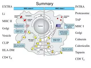
Discussion Topics • Experimental Background • Focus of the Study • SILAC Analysis • Mitochondrial Membrane Degradation • Conclusion • Future Research
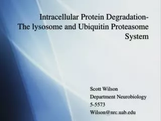
Experimental Background Parkin is a Parkinson’s disease (PD) related protein. When mitochondria malfunction Parkintranslocates from the cytosol in order to aid in degrading them via mitophagy. Parkin is an E3 ubiquitinase that attaches the protein ubiquitin to malfunctioning mitochondria in a chain linked fashion marking them for degradation. Two major pathways of Parkin mediated polyubiquitization and degradation are reported. • The ubiquitin-proteasome system (UPS), which uses the K48-linked polyubiquitization of substrate proteins, whose malfunction would lead to an accumulation of misfolded proteins (Lewy bodies) • The K63-linked polyubiquitization of mitochondrial proteins, which recruits ubiquitin adaptors of the autophagic machinery.(HDAC6 and p62/SQSTM1)
What was the study’s focus? Key Concepts • K48-mediated UPS pathway’s association with mitophagy • 26S proteasome’s involvement in mitochondrial outer membrane protein degradation by the UPS • UPS is a necessity of mitophagy and is activated prior • UPS inhibition prevents mitophagic function This study was focused on better understanding the function of Parkin and the associated changes to the mitochondrial proteome in response to Parkin.
SILAC • 2 hours of treatment with CCCP depletes mitochondrial membrane potential in Parkin-expressing HeLa S3 clones. • Parkin-dependent mitophagy results • 2979 different protein groups identified. 1013 mitochondrial proteins Stable isotope labeling by amino acid culture analysis (Mass Spec Approach)
SILAC Interpretation • Parkin increased 13 fold • P62/SQSTM1, NBR1, LC3 and GABARAPL2 autophage related proteins highly elevated • V-type proton ATPase increased (component of endosomes and lysosomes) • Drp1 had increased output (involved in mitochondrial fragmentation) • UPS system induced: 9 fold increase in ubiquitin and increased proteosome subunits 19S and 20S of the 26S proteosome • Outer membrane proteins reduced (Mfn1, Mfn2, Miro1 and Miro2) • Tom70 substantially reduced
Mitochondrial Outer Membrane Degradation Figure 1A: Immunoblotting revealed rapid degradation of Mfn1, Mfn2 and Tom70 after CCCP treatment of Parkin-expressing cells after approximately one hour VDAC1 (solute transport), Fis1 (mitochondrial fission), Bak (cellular apoptosis) and Tom20 (protein import) also reduced but over a longer time period Intermembrane and matrix proteins of mitochondria showed no significant changes Opa1 has short and long isoforms. The long isoform (outer membrane) is reduced while the short isoform (inner membrane) is nearly unchanged Figure 1B and 1C: Treatment with MG132 or epoxomicin proteosome inhibitors before CCCP treatment inhibited membrane degradation These results indicate that Parkin is activated by mitochondrial membrane depolarizations and mediates proteosome-dependent outer membrane degradation.
Mitochondrial Membrane is Degraded Independent of Autophagy Atg3 is similar to the E2 enzyme In Atg3-null MEFs do not show conjugation of AT8/ LC3 to phosphatidylethanolamine, indicating the autophagic pathway has failed to activate. The addition of epoxomicin blocked membrane degradation This indicates that Parkin degrades the outer-membrane proteins via the UPS
UPS Up Regulation Figure A: SILAC ratio results for K48 and K63-linked polyubiquitinization Figure B: Anti-polyubiquitin K48 and K63 antibody expression is prevalent in mitochondria of Parkin expressing cells upon CCCP treatment Figure C: 26S β subunits of the proteasome stained. Upon CCCP treatment the proteosame is centered upon mitochondria. Figure D: Same as Figure C only using the α subunit.
Temporal Relationship: Outer Membrane Degradation and Mitophagy Tom20 outer membrane protein is compared with Hsp60 of the mitochondrial matrix. Figure A-B: Parkin-expressing cells treated with CCCP segregated into two distinct populations. Perinuclear mitochondria were aggregated in nuclear exterior and retained both Tom20 and Hsp60. Peripheral mitochondria showed no aggregation and no change in Hsp60 but a loss off Tom20. Figure C: % of Tom20 negative cells that were Hsp60 positive after 24 hours. Figure D: Outer membrane proteins were measured in wild-type MEFs and atg-null MEFs after CCCP treatment and were shown to be degraded in a similar fashion while Hsp60 was virtually unaltered.
Parkin- Mediated Mitophagy Requires the UPS LC3B is an autophagosome marker Figure A: Dispersed mitochondria stained for LC3B show and Hsp60 Figure B: MG132 proteasome inhibitor prevents the loss of Tom20 as a result of CCCP treatment.. Figure C- D: 12 and 24 hr recovery after 100min pulse of CCCP Figure E: Parkin expressing human neuroblastoma SH-HY5Y cells treated with CCCP for 4 hours Figure F: Epoxomicin prevents mitophagy due to CCCP treatment .
Epoxomicin blocks mitophagy in CCCP treated cells EGFP marked cells used to indicating mitofusins may help in mitochondrial segregation but more proteasome remodeling, including proteolysis, is necessary for mitophagy.
Conclusion Parkin is part of a mitochondrial quality control mechanism Parkin is part of two distinct degradative mechanisms, the UPS and autophagy which are linked in mitochondrial control Outer membrane degradation by Parkin via the UPS is a signal for autophagosomes to target mitochondria Outer membrane degradation by the UPS causes dispersal of mitochondria which leads to autophagosome targeting and engulfment
Future Research Authors suggestions: Future research on mitophagy will require a matrix protein marker for accurate assessment. Investigate the UPS role in other forms of autophagy. Does Parkin mediated activation of UPS have other functions for mitochondria besides autophagy? Other questions: Why were the Atg-null mutants not tested with the antibodies for K-48 and K-63? What signals causes mitochondrial aggregation and dispersal? Tom20 degradation or another protein?