Laparoscopic Pyeloplasty by Jaime Landman, MD
190 likes | 656 Vues
Learn about the procedure, patient positioning, trocar placement, steps, technical tips, and caveats explained by Dr. Jaime Landman, a renowned urologist. Discover the required equipment and post-operative care to improve patient outcomes.

Laparoscopic Pyeloplasty by Jaime Landman, MD
E N D
Presentation Transcript
Laparoscopic Pyeloplasty Jaime Landman, MD Assistant Professor of Urology Columbia University Department of Urology
Patient Selection • Indications • Symptomatic UPJ obstruction • Asymptomatic UPJ obstruction with documented deterioration of renal function • Contraindications • Intra-renal pelvis • Multiple prior abdominal surgeries (relative)
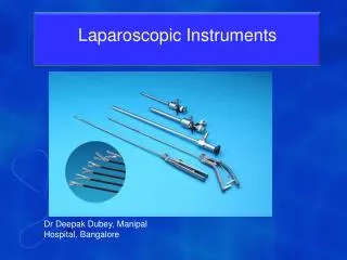
Equipment Required • Veress needle (14G) • Knife with #15 blade • Dilating trocars: 12 mm (1), 5 mm (2 or 3) • 10 mm laparoscope with 30 Lens • 5 mm laparoscope with 0 Lens • Harmonic Shears-5 mm curved [Ethicon] • Macrobipolar grasper [Aesculap] • Endoshears with disposable tip (Microline) • 5 mm suction irrigation device • Endoholder (self-retaining retractor) [Codman] • Padron Endoscopic Retractor (PEER) [J Jamner] • Suture: 4-Vicryl on an SH needle • Lapra-Ty clip applier and clips [Ethicon] • 7 mm Jackson Pratt drain
Patient Positioning • Standard full flank, ventral surface on edge of table • Lower (left) leg bent, 3 pillows supporting upper (right) leg • Table flexed 15 • Axillary roll • OR table covered with gel pad (never bean bag) • Arm draped over chest, supported by 2 pillows • Arms, hips, and lower leg secured by tape • No kidney rest
Patient Positioning = Areas that are carefully padded
Trocar Placement • Veress needle placed medial and superior to the anterior superior iliac spine, followed by 5 mm trocar that will be the right hand working site • 5 mm trocar beneath costal margin in anterior axillary line for left hand working site • 12 mm trocar in midline between the two working trocars for the laparoscope • Optional 5 mm trocar in posterior axillary line between the working trocars for lateral retraction
Port Placement =12mm trocar =5 mm trocar =5 mm trocar (optional)
Port Placement (post-operative) Feet Head 7mm Jackson Pratt drain in retractor site 5mm left hand working site 5mm right hand working site 12mm laparoscope site
Steps of the procedure • Deployment of trocars* • Mobilization of Colon and Kocherizing the duodenum (right side) • Identification and limited mobilization of ureter and the renal pelvis with preservation of crossing vessels when present *If not done pre-operatively, cystoscopy, retrograde ureteropyelogram, and JJ stent deployment can be done before laparoscopy OR a JJ stent can be deployed laparoscopically after transection of the UPJ
Steps of the procedure 4. Transection of the UPJ and spatulation of the ureter. Reduction of renal pelvis (when redundant) 5. Anastomosis (anterior to crossing vessels when present) and deployment of drain
Step 2. Mobilization of Colon and Kocherizing the duodenum (right side)
Step 3. Identification and limited mobilization of the ureter
Step 4. Transection of the UPJ and spatulation of the ureter
Technical points: Tips • Pre-operative CT angiogram is reliable in the detection of crossing vessels and can warn of the existence of crossing vessels • Patients with JJ stents placed prior to surgery will have thickened reactive ureters which may make the ureteral dissection and anastomosis more challenging • Application of the PEER retractor and the Endoholder opens the operative field and facilitates dissection • Microline scissors have a disposable tip which is always sharp for ureteral transection and spatulation • Lapra-Ty clips will securely anchor the running anterior and posterior suture lines and will facilitate a tight closure
Technical points: Caveats • Facility with intracorporeal suturing is essential and will make the running anastomosis relatively expeditious and easy • Early in the surgeon’s experience, application of three 12 mm trocars will facilitate the procedure • It is ideal to work with the laproscope between the two “working” trocars. However, the laparoscope may be moved to optimize the angle of vision • When using 5 mm working trocars, the needle and Lapra-Ty clip applier are inserted through the 12 mm (laparoscope) trocar and a 5 mm laparoscope is used
Credits Surgeon: Jaime Landman Director of Minimally Invasive Urology Columbia University Department of Urology, New York, NY Assistant: Sean Collins Director of Minimally Invasive Urology Louisiana State University Department of Urology, New Orlenes, LA