Pig Heart Dissection Workshop
Pig Heart Dissection Workshop. Pig Heart Dissection Workshop. Two parts to this session:. Pre-dissection activities:. Drawing activity Heart-Walk activity Electrophysiology activity. P ig heart dissection. Draw-the-Heart. A Pre-Dissection Pig Heart Activity.

Pig Heart Dissection Workshop
E N D
Presentation Transcript
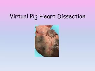
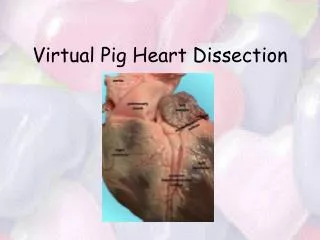
Two parts to this session: Pre-dissection activities: • Drawing activity • Heart-Walk activity • Electrophysiology activity • Pig heart dissection
Draw-the-Heart • A Pre-Dissection Pig Heart Activity D. Kacher - DeBakey High School for Health Professions
Supplies needed: • two sheets of blank paper • pencil (with eraser) • Highlighters or colored pencils (blue, red, green, yellow)
ObjectivesUpon completion of this lesson, you should be able to: • identify great vessels & valves associated with right & left sides of the heart • differentiate between systemic & pulmonary circulation • trace the pathway of blood through the heart
The heart is a double pump right heart pump left heart pump
The right heartpumps deoxygenated blood. (Called deoxygenated although it has approximately 68% - 75% oxygen.) The left heartpumpsoxygenated blood (94%-100%).
Deoxygenated blood: • called “venous” blood – leaves body tissues • returns to right heart, which pumps it to lungs where it’s “re-oxygenated” • leaves lungs and returns to left heart • Movement of blood from heart to lungs & back to heart is referred to as pulmonarycirculation or the pulmonary circuit
Step 1:Draw outline of heart; label as seen below. Rt. side Lf. side apex
Throughout this exercise, there will be yellow highlighting to show drawing sequence.
Step 2:Draw myocardium & septum; identify chambers. Rt. side Lf. side LA RA septum LV RV apex
Step 3: Draw superior vena cava (SVC) & inferior vena cava (IVC). Rt. side Lf. side SVC LA RA septum LV IVC RV apex
Step 4:Draw pulmonary artery & wall of right atrium Rt. side Lf. side SVC LA RA pulmonary artery septum LV IVC RV apex
The pulmonary artery is the only artery in the body to carry deoxygenated blood. Remember: Arteries carry blood Away from the heart.
Step 5: Draw tricuspid & pulmonary valves. Rt. side Lf. side SVC LA pulmonary artery pulmonary valve RA tricuspid valve septum LV IVC RV apex
Step 6: Outline great vessels & chambers in blue. Rt. side Lf. side SVC LA pulmonary artery pulmonary valve RA tricuspid valve septum LV RV IVC apex
Step 7: Color myocardium & valves. Rt. side Lf. side SVC LA pulmonary artery pulmonary valve RA tricuspid valve septum LV IVC RV apex
Step 8: Indicate pathway of blood with blue arrows. Rt. side Lf. side SVC LA pulmonary artery pulmonary valve RA tricuspid valve LV IVC RV apex
Left heart blood: • called “arterial” blood- leaves lungs as highly oxygenated blood (94% - 100%) • returns to left heart • pumped to organ systems (including the heart--coronary arteries) • referred to as systemiccirculation or the systemic circuit
Step 1:Draw outline of heart; label as seen below Rt. side Lf. side apex
Step 2:Draw myocardium & septum; identify chambers Rt. side Lf. side LA RA septum LV RV apex
Step 3: Draw aorta& upper wall of left atrium Rt. side Lf. side arch aortic aorta LA aortic root RA septum LV RV apex descending aorta
Step 4: Draw & label pulmonary veins Rt. side Lf. side arch aortic aorta pulmonary veins pulmonary veins LA aortic root RA septum LV RV apex descending aorta
Step 5: Draw mitral & aortic valves. Rt. side Lf. side arch aortic aorta pulmonary veins pulmonary veins LA aortic root mitral valve RA aortic valve septum LV RV apex descending aorta
“Pronunciation .” mitral = mI-trall prefix: mi- = two
Step 6: Outline chambers & great vessels in red. Rt. side Lf. side arch aortic aorta pulmonary veins pulmonary veins LA aortic root mitral valve RA aortic valve septum LV RV apex descending aorta
Step 7: Color the myocardium & valves. Rt. side Lf. side arch aortic aorta pulmonary veins pulmonary veins LA aortic root mitral valve RA aortic valve septum LV RV apex descending aorta
Step 8: Indicate pathway of blood with arrows. Rt. side Lf. side arch aortic aorta pulmonary veins pulmonary veins LA aortic root mitral valve RA aortic valve septum LV RV apex descending aorta
Post Test: • Please clear your desks. • Take out a sheet of notebook paper. • Write your name and proper heading at top of paper.
Does the left heart transport high or low oxygenated blood? • Which great vessels are associated with the right heart? • Name the heart valves associated with the right heart. • Which great vessels are associated with the left heart? • Which two heart valves are associated with the left heart? • What does the Latin word “septum” mean?
Why are heart valves “one-way” valves? • In the term mitral, what does the prefix “mi-” mean? • Which artery carries deoxygenated blood? • What does “systemic circulation” refer to? • What does “pulmonary circulation refer to?” • Which muscular wall do you think is thicker-- the right ventricular myocardium or the left ventricular myocardium– and why?