Download
1 / 33
330 likes | 486 Vues
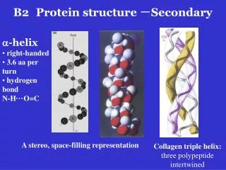
B2 Protein structure - Secondary. a -helix right-handed 3.6 aa per turn hydrogen bond N-H ··· O=C. A stereo, space-filling representation. Collagen triple helix: three polypeptide intertwined.

E N D
B2 Protein structure -Secondary • a-helix • right-handed • 3.6 aa per turn • hydrogen bond • N-H···O=C A stereo, space-filling representation Collagen triple helix: three polypeptide intertwined
b-sheet: hydrogen bonding of the peptide bond N-H and C=O groups to the complementary groups of another section of the polypeptide chainx Parallel b sheet: sections run in the same direction Antiparallel b sheet: sections run in the opposite direction A stereo, space-filling representation of the six-stranded antiparallel b sheet. fibroin
B2 Protein structure and function A two-stranded b sheet
B2 Protein structure -Tertiary The different sections of a-helix, b-sheet, other minor secondary structure and connecting loops of a polypeptide fold in three dimensions
B2 Protein structure and function Noncovalent interaction between side chains that hold the tertiary structure together: van der Waals forces, hydrogen bonds, electrostatic salt bridges, hydrophobic interactions Covalent interaction: disulfide bonds Denaturationof protein by disruption of its 2o and 3o structure will lead to a random coil conformation
B2 Protein structure -Quaternary Many proteins are composed of two or more polypeptide chains (subunits). These subunits may be identical or different. The same forces which stabilize tertiary structure hold these subunits together. This level of organization called quaternary structure. A stereo, space-filling drawing showing the quaternary structure of hemoglobin a1-yellow; b1-light blue; a2-green; b2-dark blue; heme-red back
Advantages of the quaternary structure: • It allows very large protein molecules to be made, such as tubulin。 • It can provide greater functionality to a protein by combining different activities into a single entity. • The interactions between the subunits can often be modified by binding of small molecules and lead to the allosteric effects seen in enzyme regulation
B2 Protein structure and function Protein Folding: chaperones are involved in vivo • The rapidly reversible formation of local secondary structure • Formation of domains through the cooperative aggregation of folding nuclei • Assembly of domains into a “molten” globule • Conformational adjustment of the monomer • Final conformational adjustment of the dimeric protein to form the native structure. back
B2 Protein structure and function Prosthetic groups (辅基): covalently or noncovalently attached to many conjugated proteins, and give the proteins chemical functionality. Many are co-factors in enzyme reactions. Apoprotein (脱辅基蛋白) Examples : heme groups in hemogobin (Figure)
B2 Protein structure and function • Biological functions of proteins • Enzymes: substrate binding, side chain in catalysis • Signaling: cell membrane • Transport and storage: hemoglobin transports oxygen • Structure and movement: collagen, keratin, tubulin in cytoskeleton, actin and myosin for muscle contraction • Nutrition: casein (酪蛋白) and ovalbumin(卵清蛋白) • Immunity: antibodies • Regulation: transcription factors
B2 Protein structure - Domains, motifs and families Domains: structurally independent units of many proteins, connected by sections with limited higher order structure within the same polypeptide. (Figure) They can also have specific function such as substrate binding
Structural motifs: • Groupings of secondary structural elements that frequently occur in globular proteins • Often have functional significance and represent the essential parts of binding or catalytic sites conserved among a protein family bab motif
Protein families:structurally and functionally related proteins from different sources Motif The primary structures of c-type cytochromes from different organisms
Protein purification • An essential experimental step when study any individual protein • An experimental step to purify the interested protein from other proteins and nonprotein molecules existing in the cells
The principal properties of proteins used for purification • Size: gel filtration chromatography 2. Charge: ion-exchange chromatography, isoelectric focusing electrophoresis 3. Hydrophobicity: hydrophobic interaction chromatography 4. Affinity: affinity chromatography 5. Recombinant techniques: involving DNA manipulation and making protein purification so easy
2. Charge: ion-exchange chromatography, isoelectric focusing, electrophoresis Isoelectric point (pI): the pH at which the net surface charge of a protein is zero - + - + - + - + - + - - + + - + pH=pI pH<pI pH>pI
Ion-exchange chromatography Sample mixture + + + Ion displacing Protein binding Column + anions Column + anions Column + proteins Purified protein
+ Electrophoresis Protein migrate at different position depending on their net charge
Isoelectric focusing A protein will stop moving at position corresponding to its isoelectric point (pI) in a pH gradient gel.
3. Hydrophobicity: hydrophobic interaction chromatography Similar to ion-exchange chromatography except that column material contains aromatic or aliphatic alkyl groups
4. Affinity chromatography • Enzyme-substrate binding Substrate analogs: competitive inhibitors ding • Receptor-ligandbinding d • Antibody-antigen binding
5. Recombinant techniques: • Clone the protein encodinggene of interest in an expression vector with a purification tag added at the 5’- or 3’ end of the gene • Protein overexpressionin a cell • Protein purificationwith affinity chromatography.
Mass Determination Gel filtrationchromatography and SDS-PAGE • Comparing of the unknown protein with a properstandard • PopularSDS-PAGE: cheap and easy with a 5-10% error • SDS: sodium dodecyl sulfate, makes the proteins negatively charged and the overall charge of a protein is dependent on its mass.
Mass Determination Mass spectrometry: • Molecules are vaporized and ionized (by Xe/Ar beam), and the degree of deflection (mass-dependent) of the ions in an electromagnetic field is measured • Extremely accurate (0.01% error), but expensive • ESI (electrospray ionization) and MALDI (matrix-assisted laser desorption/ionization) can measure the mass of proteins smaller than 100 KDa • Protein sequencing: relying on the protein data base • Helpful to detect post-translational modification
Determine the primary structure of a protein:p • Amino acid composition: • Acid treatment to hydrolyze peptide bonds: 6M HCl, 110°C for 24 hrs. • Chromatographic analysis • However, you cannot get the sequence!
Determine the primary structure of a protein:protein sequencing • Specific enzyme/chemical cleavage: • Pepsin cleaves after lysine(K) or arginine (R) • V8 protease cleaves after glutamic acid (E) • Cyanogen bromide cleaves after methionine (M) • Edman degradation: • Performed in an automated protein sequencer • Determine thesequence of a polypeptide from N-terminal amino acid one by one. Phenylisothiocyanate 苯异硫氰酸 reaction) • Expensive and laborious
Protein sequence analysis (1) Sequence: HLMGSHLVDALELVMGDRGFEYTPKAWLV Pepsin T1 HLMGSHLVDALELVMGDR T2 GFEYTPK T3 AWLV V8 V1 HLMGSHLVDALE V2 LVMGDRGFE V3 YTPKAWLV
Protein sequence analysis (2) Computer analysis: T1HLMGSHLVDALELVMGDR V1 HLMGSHLVDALE V2LVMGDRGFE T2GFEYTPK V3YTPKAWLV T3 AWLV Sequence: HLMGSHLVDALELVMGDRGFEYTPKAWLV
Most protein sequences are deduced from the DNA/cDNA sequence Direct sequencing: determine the N-terminal sequences or some limited internal sequence construction of an oligonulceotide or antibody probe fishing the gene or cDNA
X-ray crystallography and NMR Determing the 3-D structure of a protein X-ray crystallography: • Measuring the pattern of diffraction of a beam of X-rays as it pass through a crystal. The first hand data obtained is electron density map, the crystal structure is then deduced. • A very powerful tool in understanding protein 3-D structure • Many proteins have been crystallized and analyzed
X-ray crystallography and NMR Determing the 3-D structure of a protein NMR: Nuclear magnetic resonance (NMR) spectroscopy • Measuring the relaxation of protons after they have been excited by radio frequencies in a strong magnetic field • Measure protein structure in liquid but not in crystal • Protein measured can not be larger than 30 KDa
Summary • 20 Amino acids: D & E (-), H, K, R (+), S, T, C, N, Q (polar), G,A,V,L, I, M, P (nonpolar), F, Y,W (aromatic) • Protein structure: primary, secondary, tertiary, quaternary • Protein analysis: purification, sequencing, mass determination, X-ray crystallography and NMR