Download
1 / 15
160 likes | 328 Vues
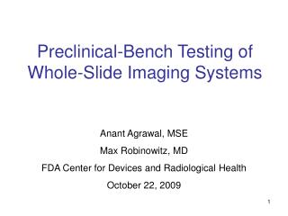
Preclinical-Bench Testing of Whole-Slide Imaging Systems. Anant Agrawal, MSE Max Robinowitz, MD FDA Center for Devices and Radiological Health October 22, 2009. Motivation. Obtain quantitative, detailed performance data under well-controlled conditions

E N D
Preclinical-Bench Testing of Whole-Slide Imaging Systems Anant Agrawal, MSE Max Robinowitz, MD FDA Center for Devices and Radiological Health October 22, 2009
Motivation • Obtain quantitative, detailed performance data under well-controlled conditions • Consistent evaluation and comparison of device performance • Reduce burden of acquiring and analyzing clinical data • Provide a straightforward means of postmarket quality control
Two Categories ofPreclinical-Bench Testing 1.Physical testing of imaging performance characteristics • Quantitative measurements of key hardware and software properties • Can isolate effects of individual components 2.Pathologist-based assessment of image quality • Using artificial phantoms and/or biological specimens (e.g., diatoms, ideal histological preparations) • To answer the question: are all critical featuresvisualized by both optical and digital microscopy systems? • Serves as a bridge to clinical studies
Preclinical-Bench Testing 1: Physical testing of imaging performance characteristics
Basic block diagram ofWhole-Slide Imaging (WSI) Conventional optical microscope SLIDE Digital image sensor Image Processing software Mechanical scanner Light source Imaging optics Display PATHOLOGIST IMAGE DATA FILES
Some imaging performance characteristics to consider:COMPONENT-LEVEL Digital image sensor Image Processing software Mechanical scanner Light source Imaging optics Display 3D Positioning accuracy Enhancement Compression Temporal stability Spatial uniformity Spectral output Color gamut Color accuracy Luminance Resolution/contrast Spatial uniformity Noise Linearity Dynamic range Spatial resolution Spatial uniformity Magnification Resolution/contrast Distortion Depth of field
Some imaging performance characteristics to consider:SYSTEM-LEVEL Digital image sensor Image Processing software Mechanical scanner Light source Imaging optics Display • Resolution and contrast • Color gamut/accuracy • Dynamic range • Spatial uniformity
Analysis software Typical paradigms forphysical system testing TEST TARGET Digital image sensor Image Processing software Mechanical scanner Light source Imaging optics Display IMAGE DATA FILES FIGURE OF MERIT (quantitative measure of performance)
Typical paradigms forphysical system testing TEST TARGET Digital image sensor Image Processing software Mechanical scanner Light source Imaging optics Display LIGHT MEASUREMENT INSTRUMENT FIGURE OF MERIT
Obtaining statistics during physical system testing • WITHIN-SYSTEM VARIABILITY • Insert test target x times into same WSI unit • BETWEEN-SYSTEM VARIABILITY • Insert test target into x different WSI units of the same model
Examples of readily-available test targets USAF 1951 resolution target Distortion and scanning accuracy grid target Modulation/ Contrast transfer function
Quantitative characterization of a typical slide image From Kayser et al, Diagnostic Pathology, July 2008.
Information needed with each measured characteristic • Test target and procedure • Quantitative figure of merit • Benchmark or specification • Compare to conventional optical microscope • Quality control schedule • Certain components (light source, mechanical scanners) may require frequent QC checks/calibration
Potential resources for testing methodology: standards • Microscope-specific • DIN 58959 (1997) – Quality management in medical microbiology - Part 4: Requirements for investigations using light microscopes • ISO 8039:1997 – Optics and optical instruments – Microscopes – Magnification • Digital image sensor • ISO 15739:2003 – Photography – Electronic still-picture imaging – Noise measurements • ISO 12233:2000 – Photography – Electronic still-picture imaging – Camera resolution • IEEE Std 208-1995 (R2005) – Measurement of Resolution of Camera Systems • Display • AAPM TG#18 “Assessment of Display Performance for Medical Imaging Systems” • IEC 62B WG36 “Image display devices” approved as of Sept 2009 • VESA “Flat Panel Display Measurements Standard”
How to Establish Physical Testing Guidelines? • Existing standards provide some testing methodology relevant to whole-slide imaging systems for histopathology • Which physical tests are the most useful to validate device performance without excessive burden to the sponsor? • System-level tests are most straightforward • Component-level testing permits easier replacement of components as system configurations evolve • Need to consider physical testing for variability within and between systems