Download
1 / 43
451 likes | 1.8k Vues
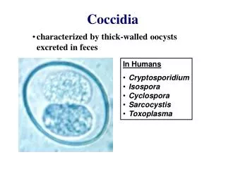
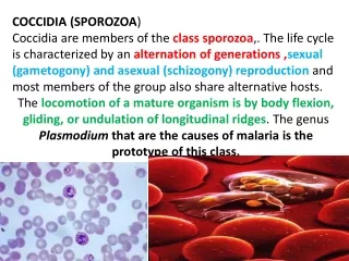
Coccidia and other Protozoa of Food Animals. Coccidia or Ruminants. Common coccidian oocysts of cattle. E bovis 28 x 20u. E zuernii 18 x 15 u. Coccidiosis: Eimeria in ruminants, poultry; Isospora in dog, cat, pig Species specific Self limiting

E N D
Common coccidian oocysts of cattle. E bovis 28 x 20u E zuernii 18 x 15 u • Coccidiosis:Eimeria in ruminants, poultry; Isospora in dog, cat, pig • Species specific • Self limiting • Contagious, especially crowding, moist transmission foci (water holes, leaky troughs) • Non-immune, young animals; ‘Multipier’ effect at exposure Only E bovis, E zuernii are definite pathogens in cattle
Eimeria oocyst Eimeria has four sporocysts with two sporozoites Isospora has two sporocysts with four sporozoites
Eimeria bovis has characteristic, 1st generation ‘giant schizonts’ in small intestine
Gross pathology – giant schizonts of E. bovis are grossly visible
The major pathogenesis of both Eimeria bovis and E zuernii is due to the sexual stages that occur deep in the mucosa of the caecum, large intestine and rectum Developing Oocyst Oocyst
Feedlot– Clinical coccidiosis is a major problem in the dairy and feedlot industries
E. bovis - clinical signs in young dairy heifer Bloody, mucoid diarrhea is often seen 1-3 days before 1st oocysts are shed. Prepatent period 16-17 days, peak oocyst production at 3 weeks. Patent period is 2-3 weeks, then self limits.
Clinical Signs • Bloody, mucoid diarrhea: +/- epithelial mucosal tags, dehydration, depression, tenesmus, occasional rectal prolapse • Seasonality: Disease most common in fall, least summer; a highly pathogenic form of the disease ‘winter coccidiosis’ of unclear epidemiology occurs, especially in feedlots above the 40th parallel • Acute death by 5-7 days: Others develop secondary enteritis, pneumonia, a few linger in poor condition and are culled • Nervous coccidiosis: Seen in a variable % of animals in outbreaks - high mortality, diarrhea, tremors, convulsions, nystagmus, blind, aggressiveness. Seen especially with some strains of E zuernii
Diagnosis: Gross pathology findings in colon and rectum. Evidence of hemorrhagic enteritis with frank blood, mucous
Fresh mucosal impression smear with coccidian developmental stages. Necropsy touch preparations or rectal scrapings can be used to diagnose prepatent infections
Treatment Drug Dosage Class Amprolium Pv 5 mg/kg for 21 days Thiamine Rx 10 mg/kg for 5 days antagonist Decoquinate Pv 0.5 mg/kg for 28 days Quarternary Ammonium Sulfaquinoxaline Rx 8-70 mg/kg per day Sulfonamide Monensin Pv 3 mg/kg/day Ionophore antibiotic Lasalocid Pv 3 mg/kg/day Ionophore antibiotic
Control • Treat to prevent incubating new cases and reduce oocyst shedding ‘multiplier effect’; clinical signs are seen after damage is done • Treat until infections self-limit and/or immunity builds • Oocysts live 1 year at 4o C, resist mild freezes; sunlight and dry heat kills oocysts in 4 hours • Provide clean dry conditions, reduce stress, crowding (eg. portable calf sheds, fix leaks) • Do not feed off ground or follow probable heavy contamination
Coccidia of sheep and goats • Most transmission in spring at lambing • 2-8 week-old lambs in confinement, 2-3 weeks post weaning or stress (eg. shipping, change of feed) • Heavily stocked irrigated pastures, contaminated wet areas, feedlot • Sheep and goats tend to have watery diarrhea, seldom bloody • Diagnosis: Several thousand oocysts/gm; most shed high oocyst ‘background’
Coccidia of sheep and goats Species Prepatent period Clinical Effects E. ahsata – Sh 18-21 days Sh +++, G + E. christenseni – G (28 x 18u) E ovina - Sh 19-29 days Sh +, G ++ E arloingi - G (28 x 20u) E ovinoidalis - Sh 9-15 days Sh +++, G +++ E ninakohlyakimovae - G (23 x 18u)
Toxoplasma gondii • Ruminant-cat cycle; oocysts can survive 18 months if cool, wet • Sheep and goats - Seldom acute disease, outbreaks of late abortion, weak lambs, pyometra, • Cattle minor; more resistant • Immunity – no additional abortions or clinical signs if re-exposed Sporulated oocysts
Diagnosis: • Placental lesions - cotyledons inflammed, greyish white foci 1-3mm (below). Impression smears reveal typical ‘banana-shaped’ organisms • Paired titers: IFA, IHA, CF, ELISA, Sabin-Feldman dye test, mouse inoculation intraperitoneal organisms Treatment: Sulfadiazine+Pyrimethimine seldom given, impractical Vaccine in New Zealand
Neospora caninum A transplacentally transmitted protozoan parasite of livestock and dogs that was until 1988 often misdiagnosed as T gondii. Dogs were shown to be a definitive host in 1998.
Abortion in dairy cattle • Retrospective studies in California specimen archives show Neospora was the cause of 18-19% of all aborted fetus tissues, with 24.4% of those from the dairy industry. It is now known to be the most common cause of bovine abortion in California, the Netherlands and New Zealand and has a worldwide distribution. • It was first recognized in dogs in Norway (1984) in association with encephalitis and myositis. Infections have been demonstrated in cattle, sheep, goats, horses, deer, dogs (which serve an intermediate and definitive host), cats and mice. • Abortion is also reported naturally for goats, experimentally for sheep.
Clinical signs and pathogenesis • Year-round abortion in heifers and cows, mainly at 3-6 months • Intensive management favors transmission. Contamination of feedstuffs with dog feces apparently quite common; Sarcocystis bovicanis is ubiquitous in cattle. Dairy and beef equally susceptible • Repeat abortions can occur repeatedly in the same herd (~5%, up to 30%) and the same animal, unknown it re-infection or reactivation • Congenitally infected calves have CNS damage ranging from proprioceptor deficits to paralysis or no signs but high pre-colostral antibody titers. Vertical congenital transmission, with abortion in offspring is documented. This broad range of effects is possibly related to infective dose or to strain of parasite.
Diagnosis and control • Submit aborted calf, placenta and serum from dam • Histopathology shows non-suppurative multifocal protozoal encephalomyelitis, especially in spinal grey matter, and myocarditis, portal hepatitis and lesions in cardiac and skeletal muscles. Neospora has thicker walled cysts than Toxoplasma • Immunohistochemistry (IFAT) of tissue sections, ultrastructure or species specific PCR differentiates Neospora from Toxoplasma or Sarcocystis • ELISA and IFAT (>1:640) is useful for sero-epidemiology evaluations. Risk of seropositive cows aborting is twice that of negative titer, possibly by congenital exposure from dam. • Limit access of young dogs. Litters may develop fatal ascending paralysis, myocarditis with dermal lesions. Variable treatment success with sulfadiazine/pyrimethemine, clindamycin.
Sarcocystis • A Predator-Prey life cycle. Many highly host-specific species occur • A large number of fully sporulated sporocysts are shed by predators
Sarcocystis in muscle Sarcocystis bovicanis – Cause of uncommon acute outbreaks of ‘abortion storm’ (Dalmeny disease). Signs are fever, anorhexia, cachexia, abortion, some deaths, characteristic ‘rat tail’ associated with 2 asexual reproduction generations in endothelial cells of highly vascular organs. Muscle ‘sarcocysts seen above are 3rd generation schizonts, a frequent gross pathological finding in cardiac, esophageal muscles of slaughter cattle.
Cryptosporidium Autogenous re-infection cycle by Type1 thin walled cysts (20%); Thick walled Type two cysts pass in feces to the environment.
Cryptosporidium in superficial mucosa Causes villous atrophy; mononuclear infiltrate in lamina propria, especially in the ileum; mucofibrinous exudate in severe cases.
Cryptospridium –histopathology section, H&E Cysts appear to be on cells, but are just under cell membrane at microvillous border. Immediate fixation or lose by autolysis.
Clinical signs and epidemiology • C. parvum (6 x 4u) occurs mainly in neonatal to <4 week-old calves as a part of the calf scours complex, in most cases concurrent with rotavirus, coronavirus and/or E. coli. • Yellow, loose diarrhea of 2 weeks duration, 4-day prepatent period • Immunity limits life cycle, but autoinfection persists if immuno-suppressed. No treatment, nitazoxinide effective experimentally. • Low host specificity. Calf-human transmission is well documented and can be fatal in AIDS patients. Recent evidence from CDC community outbreaks in water supplies and use of PCR reveals two strains that infect humans, Genotype 1 (human-human) and Genotype 2 (calf-human). Most outbreaks were due to sewage contamination vs agricultural waste runoff. Cryptosporidium can be found in most natural waters, but much may be strains or species (eg C. muris (rodents), C. agilis (birds) of questionable infectivity.
Cryptosporidium –Oocysts, Phase Contrast Microscopy after Sheather’s sugar flotation. Note the typical ‘dot’ (residual body). Up to millions per gram of feces are shed.
Cryptosporidium - acid fast stain; Cryptosporidium stains bright pink against a green background. Differentiate from commonly found yeasts, which are not acid-fast, are variable in size and occasionally have ‘budding’ reproductive forms.
Giardia duodenalis Cysts 12 x 8u Trophozoite • Recently shown to be an underestimated cause of diarrhea in calves in dairies, feedlots. Less prevalent in lambs. • Resembles Cryptosporidium clinically. Giardia is less age dependent and signs with shedding persists for 2-24 weeks • Rx - Fenbendazole (5 mg/kg) at normal anthelminthic dose
Tritrichomonasfoetus Note the 3 anterior flagellae and an undulating membrane
Bovine Trichomoniasis • Venereally transmitted cause of early ‘silent’ abortion that can lead to a 15 % lower calf crop, prolonged calving interval (100-day delay in conception) and occasional pyometra • Establishes in crypts of preputial fornix of bulls. No effect on semen quality, libido or excess preputial discharge. Direct correlation between age and infection rates. Bulls <2-years old are somewhat refractory; If over 3 years old, bulls become and remain infected for life due to the development of deeper, more suitable crypts in the preputial epithelium. • Common on shared, open ranges with co-mingled herds. In the western USA surveys show 5-8% of bulls are infected; a 1990 survey in CA revealed 16% of herds and 27% of bulls infected. Common on LA coastal marsh, other southern states. Transmission rate high (42%) by natural breeding, AI possible.
Pathogenesis • Colonizes vagina, uterus and oviducts after coitus. Pregnancy is established, but fetus loses viability via cytotoxic effect on placentomal interface by 50-70 days. Since there is maternal recognition of pregnancy at 14-18 days, 21-day estrus cycles are skipped. In dairies, cows test pregnant at early palpation, then abort or resorb. • Cows self-cure by 3rd heat cycle, then are immune and can be successfully re-bred. Mild vaginitis and cervicitis occurs with mild discharge. • If the fetus is not expelled, the corpus luteum is retained and pyometra may develop (up to 10% of cows) with fluid teeming with organisms. The cow becomes a permanent carrier.
Diagnosis Bulls: Sample and culture smegma from preputial cavity at fornix level by scraping using an insemination pipette. Incubate at 37C for up to 4 days, with daily examination for organism with typical jerky, rotating motility and morphology. Trypticase-yeast extract-maltose (TYM) or Diamond’s media (1% agar added) used previously. Commercial ‘InPouch TF kits (Biomed, Diagnostics, San Jose, CA) has a 2 chamber plastic pouch with proprietary media. The upper chamber is inoculated (for immediate exam), then squeezed into lower chamber for culture. Kit has less technical error, convenience, longer shelf life and is 80-90% accurate on a single culture, 96-99% on two and <1% on three. Multiple bull examination improves ‘herd diagnosis accuracy of single samples. Saline lavage less accurate Cows: Culture of females is 50-60% accurate. Pyometra fluid is teeming with organisms.
Pentatrichomonas spp Fecal contaminant species sometimes colonize the prepuce. Differentiation is possible by morphology (4-5 anterior flagellae), slow culture growth or an experimental PCR test for Tritrichomonas foetus. Special stains (DifQuik+ Lugol’s iodine), scanning electron microscopy, and PCR may be done in questionable cases on valuable animals, especially young bulls.
Tritrichomonas - Scanning EM Note the three anterior flagellae and the flagellae associated with the undulating membrane running posteriorly
Treatment and control • Herd Health Program if risk factors (prior diagnosis, old bulls, fences) • Culture all bulls at pre-season breeding soundness examination +/- after season; 3 consecutive herd examinations before deem individuals uninfected; cull infected. Metrinidazole, Imidazole IV treatment partly effective, but declared ILLEGAL by the FDA. Topical Rx ineffective. • Use young <2 year-old preferably virgin bulls that are culture negative. • Vaccinate (Trich Guard, Fort Dodge) cow herd twice (one month apart) for 2 weeks before breeding season. Killed whole organism inhibits ability of organism to adhere to vaginal epithelium, provide partial cow herd protection but no efficacy for bulls. • Segregate cow herd after palpation: Pregnant > 5 months (safe), pregnant < 5 months (cull if abort), Open (if pyometra, cull). • Include neighbor in program or have good fences.