1 / 0

Animals Eat Food

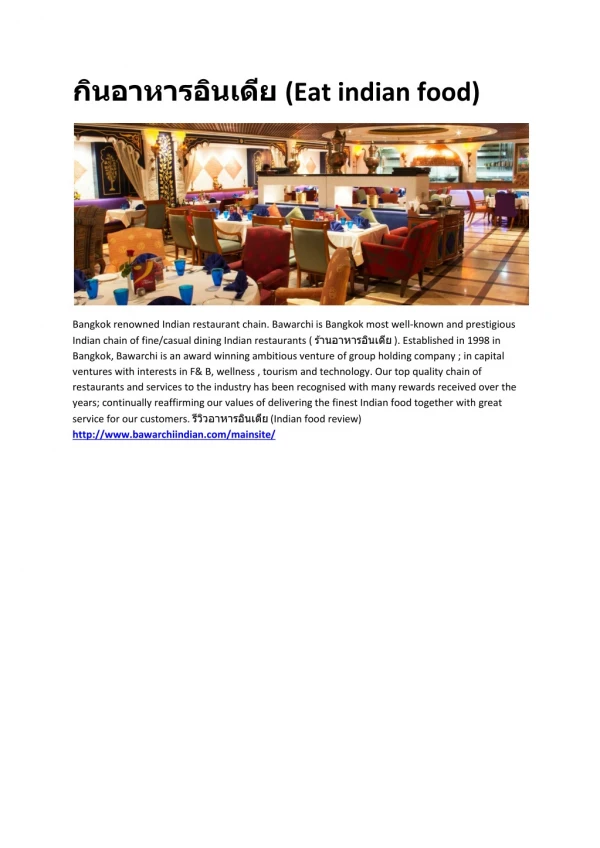
Animals Eat Food. Chap 41 (nutrition). Overview: The Need to Feed. Food is taken in, taken apart, and taken up in the process of animal nutrition In general, animals fall into three categories: Herbivores eat mainly autotrophs (plants and algae) Carnivores eat other animals

presley
Télécharger la présentation

Animals Eat Food

An Image/Link below is provided (as is) to download presentation Download Policy: Content on the Website is provided to you AS IS for your information and personal use and may not be sold / licensed / shared on other websites without getting consent from its author. Content is provided to you AS IS for your information and personal use only. Download presentation by click this link. While downloading, if for some reason you are not able to download a presentation, the publisher may have deleted the file from their server. During download, if you can't get a presentation, the file might be deleted by the publisher.

E N D

Presentation Transcript


  1. Animals Eat Food

    Chap 41 (nutrition)
  2. Overview: The Need to Feed Food is taken in, taken apart, and taken up in the process of animal nutrition In general, animals fall into three categories: Herbivores eat mainly autotrophs (plants and algae) Carnivores eat other animals Omnivores regularly consume animals as well as plants or algal matter
  3. Fig. 41-1
  4. Concept 41.1: An animal’s diet must supply chemical energy, organic molecules, and essential nutrients An animal’s diet provides chemical energy, which is converted into ATP and powers processes in the body Animals need a source of organic carbon and organic nitrogen in order to construct organic molecules Essential nutrients are required by cells and must be obtained from dietary sources
  5. Essential Nutrients There are four classes of essential nutrients: Essential amino acids Essential fatty acids Vitamins Minerals
  6. Fig. 41-2 Essential amino acids for adults Beans and otherlegumes Methionine Valine Threonine Phenylalanine Leucine Corn (maize)and other grains Isoleucine Tryptophan Lysine
  7. Fig. 41-3
  8. Vitamins Vitamins are organic molecules required in the diet in small amounts 13 vitamins essential to humans have been identified Vitamins are grouped into two categories: fat-soluble and water-soluble
  9. Table 41-1
  10. Table 41-2
  11. Undernourishment An undernourished individual will Use up stored fat and carbohydrates Break down its own proteins Lose muscle mass Suffer protein deficiency of the brain Die or suffer irreversible damage Malnourishment can cause deformities, disease, and death Malnourishment can be corrected by changes to a diet
  12. Fig. 41-4
  13. Suspension Feeders Many aquatic animals are suspension feeders, which sift small food particles from the water
  14. Fig. 41-6a Baleen Humpback whale, a suspension feeder
  15. Substrate Feeders Substrate feeders are animals that live in or on their food source
  16. Fig. 41-6b Leaf miner caterpillar,a substrate feeder Caterpillar Feces
  17. Fluid Feeders Fluid feeders suck nutrient-rich fluid from a living host
  18. Fig. 41-6c Mosquito, a fluid feeder
  19. Bulk Feeders Bulk feeders eat relatively large pieces of food
  20. Fig. 41-6d Rock python, a bulk feeder
  21. Digestion is the process of breaking food down into molecules small enough to absorb In chemical digestion, the process of enzymatic hydrolysis splits bonds in molecules with the addition of water Absorption is uptake of nutrients by body cells Elimination is the passage of undigested material out of the digestive compartment
  22. Fig. 41-7 Smallmolecules Piecesof food Chemical digestion(enzymatic hydrolysis) Nutrientmoleculesenter bodycells Mechanicaldigestion Undigestedmaterial Food Ingestion Digestion Elimination Absorption 2 4 1 3
  23. Digestion types In intracellular digestion, food particles are engulfed by endocytosis and digested within food vacuoles Extracellular digestion is the breakdown of food particles outside of cells It occurs in compartments that are continuous with the outside of the animal’s body
  24. Fig. 41-8 Tentacles Gastrovascularcavity Food Mouth Epidermis Gastrodermis
  25. Animals with simple body plans have a gastrovascular cavity that functions in both digestion and distribution of nutrients More complex animals have a digestive tube with two openings, a mouth and an anus This digestive tube is called a complete digestive tract or an alimentary canal It can have specialized regions that carry out digestion and absorption in a stepwise fashion
  26. Fig. 41-9a Crop Gizzard Intestine Esophagus Pharynx Anus Mouth Typhlosole Lumen of intestine (a) Earthworm
  27. Fig. 41-9c Stomach Gizzard Intestine Mouth Esophagus Crop Anus (c) Bird
  28. Concept 41.3: Organs specialized for sequential stages of food processing form the mammalian digestive system The mammalian digestive system consists of an alimentary canal and accessory glands that secrete digestive juices through ducts Mammalian accessory glands are the salivary glands, the pancreas, the liver, and the gallbladder Food is pushed along by peristalsis, rhythmic contractions of muscles in the wall of the canal Valves called sphincters regulate the movement of material between compartments
  29. Fig. 41-10a Tongue Sphincter Oral cavity Salivary glands Pharynx Esophagus Sphincter Liver Stomach Ascendingportion oflarge intestine Gall-bladder Duodenum ofsmall intestine Pancreas Smallintestine Smallintestine Largeintestine Rectum Anus Appendix Cecum
  30. Fig. 41-10b Salivaryglands Mouth Esophagus Gall-bladder Stomach Smallintestine Liver Pancreas Largeintestine Rectum Anus A schematic diagram of thehuman digestive system
  31. The Oral Cavity, Pharynx, and Esophagus The first stage of digestion is mechanical and takes place in the oral cavity Salivary glands deliver saliva to lubricate food Teeth chew food into smaller particles that are exposed to salivary amylase, initiating breakdown of glucose polymers
  32. The tongue shapes food into a bolus and provides help with swallowing The region we call our throat is the pharynx, a junction that opens to both the esophagus and the trachea (windpipe) The trachea leads to the lungs The esophagus conducts food from the pharynx down to the stomach by peristalsis Swallowing causes the epiglottis to block entry to the trachea, and the bolus is guided by the larynx, the upper part of the respiratory tract Coughing occurs when the swallowing reflex fails and food or liquids reach the windpipe
  33. Fig. 41-11-3 Food Epiglottisup Tongue Epiglottisup Pharynx Esophagealsphinctercontracted Epiglottisdown Glottis Glottisdownand open Esophagealsphinctercontracted Larynx Trachea Esophagus Esophagealsphincterrelaxed Glottis upand closed Relaxedmuscles Tolungs Tostomach Contractedmuscles Relaxedmuscles Sphincterrelaxed Stomach
  34. Digestion in the Stomach The stomach stores food and secretes gastric juice, which converts a meal to acid chyme The esophagus conducts food from the pharynx down to the stomach by peristalsis Swallowing causes the epiglottis to block entry to the trachea, and the bolus is guided by the larynx, the upper part of the respiratory tract Coughing occurs when the swallowing reflex fails and food or liquids reach the windpipe
  35. Fig. 41-12 Esophagus Sphincter Stomach Sphincter 5 µm Small intestine Folds ofepithelialtissue Interior surfaceof stomach Epithelium 3 Pepsinogen and HClare secreted. 1 Pepsinogen Pepsin 2 HCl Gastric gland 2 HCl convertspepsinogen to pepsin. 1 3 Pepsin activatesmore pepsinogen. Mucus cells H+ Cl– Chief cells Chief cell Parietal cells Parietal cell
  36. Digestion in the Small Intestine The small intestine is the longest section of the alimentary canal It is the major organ of digestion and absorption
  37. Pancreatic Secretions The pancreas produces proteases trypsin and chymotrypsin, protein-digesting enzymes that are activated after entering the duodenum Its solution is alkaline and neutralizes the acidic chyme
  38. Bile Production by the Liver In the small intestine, bile aids in digestion and absorption of fats Bile is made in the liver and stored in the gallbladder
  39. Secretions of the Small Intestine The epithelial lining of the duodenum, called the brush border, produces several digestive enzymes Enzymatic digestion is completed as peristalsis moves the chyme and digestive juices along the small intestine Most digestion occurs in the duodenum; the jejunum and ileum function mainly in absorption of nutrients and water
  40. Absorption in the Small Intestine The small intestine has a huge surface area, due to villiand microvillithat are exposed to the intestinal lumen The enormous microvillar surface greatly increases the rate of nutrient absorption
  41. Fig. 41-15 Microvilli (brushborder) at apical(lumenal) surface Vein carrying bloodto hepatic portal vein Lumen Bloodcapillaries Epithelialcells Basal surface Muscle layers Largecircularfolds Epithelial cells Villi Lacteal Key Lymphvessel Nutrientabsorption Villi Intestinal wall
  42. Each villus contains a network of blood vessels and a small lymphatic vessel called a lacteal After glycerol and fatty acids are absorbed by epithelial cells, they are recombined into fats within these cells These fats are mixed with cholesterol and coated with protein, forming molecules called chylomicrons, which are transported into lacteals
  43. Absorption in the Large Intestine The colon of the large intestine is connected to the small intestine The cecumaids in the fermentation of plant material and connects where the small and large intestines meet The human cecum has an extension called the appendix,which plays a very minor role in immunity
  44. Fig. 41-17
  45. A major function of the colon is to recover water that has entered the alimentary canal Wastes of the digestive tract, the feces, become more solid as they move through the colon Feces pass through the rectum and exit via the anus
  46. The colon houses strains of the bacterium Escherichia coli, some of which produce vitamins Feces are stored in the rectum until they can be eliminated Two sphincters between the rectum and anus control bowel movements
  47. Fig. 41-18 Incisors Molars Canines Premolars (a) Carnivore (b) Herbivore (c) Omnivore
  48. Fig. 41-19 Small intestine Stomach Small intestine Cecum Colon(largeintestine) Carnivore Herbivore
  49. Fig. 41-20 Rumen Reticulum 1 2 Intestine Esophagus Abomasum Omasum 4 3
  50. Overnourishment and Obesity Overnourishment causes obesity, which results from excessive intake of food energy with the excess stored as fat Obesity contributes to diabetes (type 2), cancer of the colon and breasts, heart attacks, and strokes
  51. Fig. 41-24a EXPERIMENT Obese mouse with mutantob gene (left) next to wild-type mouse.
More Related