RESPIRATION PRACTICALS
RESPIRATION PRACTICALS. SPIROMETRY. by Dr . Thouraya BOUTKEDIIRT. Objectives. Use a spirometer and determine lung volumes and capacities Define and provide values for the various lung volumes and capacities
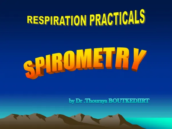

RESPIRATION PRACTICALS
E N D
Presentation Transcript
RESPIRATION PRACTICALS SPIROMETRY by Dr .Thouraya BOUTKEDIIRT
Objectives Use a spirometer and determine lung volumes and capacities Define and provide values for the various lung volumes and capacities Recognize the physiological and some pathological factors that modify lung volumes and capacities
What is spirometry ? Spirometry is a pulmonary function test that measures lung volumes and capacities
Why a spirometry test ? • To determine the cause of shortness of breath. • To rule out any kind of obstructive or restrictive disease. • To diagnose and monitor lung problems. • To monitor how well medications for lung problems are working .
Simple Spirometer Drum inverted over a chamber of water with the drum counterbalanced by a weight In the drum is air or Oxygen A Tube connects the mouth with the gas chamber When one breathes in and out of the chamber, the drum rises and falls and an appropriate recording is made on a moving paper
Method • Insert a sterilized mouthpiece • Close the nose with the nose clip • Take a normal breath through the mouthpiece for a short time then take a deep inspiration to fill the lungs completely, then breathe normally for a short time. • Expire, forcibly as completely as possible, then breathe normally for a short time. • Take a deep forceful inspiration and immediately expire forcibly and as completely as possible, then breathe normally. • The spirogram is recorded on a moving drum
Inspiratory reserve volume Inspiratory capacity Total lung capacity Tidal volume Expiratory reserve volume Vital capacity Residual volume
The air in the lungs can be subdivided on this diagram into 4 volumes and 4 capacities:
Lung volumes • Tidal volume: The amount of air that moves into the lungs with each inspiration (or the amount that moves out with each expiration) = 500ml. • Inspiratory reserve volume: The extra volume of air that can be inspired above the normal tidal volume = 3000ml
Expiratory reserve volume: The volume expelled by an active expiratory effort after passive expiration ( after the end of a normal tidal expiration) = 1100ml • Residual volume: The air left in the lungs after the most forceful expiration = 1200ml
Lung Capacities • The inspiratory capacity : IC= TV+IRV The amount of air a person can breathe beginning at the normal expiratory level and distending the lungs to the maximum amount = 3500ml • The functional residual capacity : FRC= ERV+RV The amount of air that remains in the lungs at the end of normal expiration =2300ml
The vital capacity: VC= IRV+TV+ERV The maximum amount of air a person can expel after maximal inspiration = 4600ml • The total lung capacity: TLC= VC+RV The max volume to which the lungs can be expanded with the greatest possible inspiratory effort = 5800ml
Physiological factors influencing lung volumes and capacities • Sex: female 20-25% less • Age: ↓ VC • Obesity: ↓ VC • Height: ↑VC • Athletes: ↑ VC
Pathological factors Vital capacity is decreased with : • ↓lung volume: eg: surgical removal of lung tissues large tumors • Restrictive lung disease: inability to fully expand the lungs. eg: Pneumonia, pulmonary edema, broken ribs • Obstructive lung disease eg: Chronic bronchitis, asthma, foreign body • Loss of elastic recoil eg: emphysema