Retina Identification
Retina Identification. BIOM 426: Biometrics Systems. Instructor: Natalia Schmid. Outline. Anatomy Recognition System based on Retina Processing Measure of Performance Results Pros and Cons References . Introduction. What is Retinal Identification (RI)?

Retina Identification
E N D
Presentation Transcript
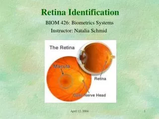
Retina Identification BIOM 426: Biometrics Systems Instructor: Natalia Schmid April 12, 2004
Outline • Anatomy • Recognition System based on Retina • Processing • Measure of Performance • Results • Pros and Cons • References April 12, 2004
Introduction • What is Retinal Identification (RI)? • Applications: very high security environments - nuclear research weapon sites, - communications control facilities, and - very large transaction processing centers. • Small user base because of: - historically high price, - unfriendly perception. April 12, 2004
History Drs. Carleton Simon and Isodore Goldstein (1935) Every eye has its own totally unique pattern. C. Simon and I. Goldstein, “A new Scientific Method of Identification,” New York State Journal of Medicine, vol. 35, no. 18, pp. 901 – 906, Sept. 1935. Dr. Paul Tower (1950’s) Studied Identical twins. Of all compared factors, retinal patterns showed the least similarity The eye shares the same stable environment as a brain and among physical features is unique to Individuals. Retinal vasculature is very stable. It’s an internal well protected from environment body (contrary to fingerprint or palm print, etc.) April 12, 2004
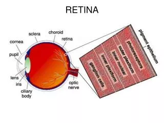
Anatomy of Eye April 12, 2004
Retina/Choroid • Retina detects incident light in the form of an image focused by a lens. • It is an internal part of the eye (well protected). • Blood comes through vessels over the Optical nerve. http://www.stlukeseye.com/anatomy/Retina.asp April 12, 2004
Retina/Choroid • Choroidal vasculature is a matting behind the retina. • Infrared illumination (retina is transparent to it). • Retinal Identification = Choroid identification = Eye Fundus (medical term) April 12, 2004
Background • EyeDentify, Inc. in 1976 began research and development of RI system. • - Late 70’s: several brands of fundus cameras were modified to obtain live image of retina for identification purpose. • - Disadvantages: 1. alignment required an assistance of operator; • 2. bright illumination was necessary (visible light was used); • 3. too complex and expensive. • First practical working prototype of RI was build in 1981. • Near-infrared light; • desktop computer for analyzing reflected waveforms; • a number of feature extraction algorithms were evaluated; • simple correlation worked best. April 12, 2004
First RI System • Product EyeDentifcation System 7.5 (4years later). • It performed: enrollment; verification; recognition. • Used circular fundus image composed of 256 twelve bit logarithmic samples. • The contrast pattern was coded in frequency domain. • Additional 32 bytes per eye of time-domain information was added to speed up the Recognition mode. • Nine active US patents. April 12, 2004
Technology • Three major subsystems of RI system are: • Imaging, signal acquisition, and signal processing: An RI camera that translates a circular scan of the retina/choroid into a digital form. • Matching: A computer that verifies or recognizes the acquired eye pattern with a stored template. • Representation: The eye (retina) signature reference templates with the corresponding identification information; storage issues. April 12, 2004
Template - Image: circular spot. - Size is chosen to return sufficient amount of light. Two major representations are: - Frequency domain; 40 bytes of contrast information; real and imaginary coordinates are encoded. - Time domain; 48 bytes of contrast information; computationally efficient. Brightness of circular spot is normalized: - brightness at a point over the average brightness. April 12, 2004
Template Eye signature = normalized contrast waveform of scan circle. Example: - 96 four-bit numbers for each of 96 equally spaced scan circle positions. April 12, 2004
RI Camera • Retinascope (medical device) - Light source is projected on subjects retina; - Returned light is detected; - Light is a collimated beam; - Due to lens light leaves eye under the same angle (retro-reflection) - In RI light detector replaces doctor’s eye April 12, 2004
Old Camera - Aligning/Fixation : (33) fixation target allows user to correctly focus and align his visual axis (10) with the optical axis (34). Allign ghosty spots. - Scan is initiated. - An IR source (39) generates a beam. An IR filter (46) stops visible light. (52) splits the beam. - The scanner directs light from 10 degree angle. A rotatable housing (57) rotates the 10 degree offset beam. - Reflected from fundus (12) light is re-collimated by the lens (30) back to the objective lens (66). April 12, 2004
Signal Acquisition Subsystem Light from RI camera Detector/ Preamplifier Op-amplifier Op-amplifier voltage Signal converter Amplifies signal A/D converter Contrast Processor Digitizes signal Removes redundant information from the waveform. April 12, 2004
System Operation To take eye reading: 1. Take off glasses (if you wear). 2. Enter PIN (verification system). 3. Position camera at eye level. 4. Align dots . The smaller dot will appear inside a larger dot. 5. Both eyes should be wide open. 6. Distance between the eye lens and eye should be < 3/4 inch. 7. Press scan button. 8. Hold your head steady during the reading. Aligment is critial. RI at a distance: Working RI systems with an operating distance 12 inch were demonstrated in the laboratory. April 12, 2004
Performance Approximately Gaussian with mean 0.144 and std 0.117. Mismatch Frequency Distribution Matching: 1.Signature is sampled 2. Normalization to have RMS value of 1.0. 3. Correlation Matching (1 is a perfect match; -1 is a perfect mismatch). Threshold is t = 0.7. The FAR is one in one million. April 12, 2004
Source of Errors False reject can be caused: • Lack of fixation • Incorrect eye distance to RI camera lens • Insufficient pupil size • Obstruction and distortion of the optical path from: - dirty camera window - contact lens edges - subject neglects to remove glasses • Ambient light interference Small pupil can cause False Reject. The amount of reflected light is inversely proportional to d^4 (d is the pupil size). April 12, 2004
Limitations • Perceived Health Threat • Oudoors vs. Indoors (noise is larger than signature) • Ergonomics: more practical in”workstation” applications while the oposite is true in physical access control applications. • Severe Astigmatism (difficulty to align dots). • High camera cost (puts lower limit on cost of RI system). April 12, 2004
Future RI systems provide a high accuracy E-commerce applications: RI might reach a critical mass. With small size signature and high accuracy RI provides better encryption tool than cryptography that depends on a key. April 12, 2004
References 1. A. Jain et al. Biometrics: Personal Identification in Networked Society, Kluwer, 1999. 2. R. B. Hill, “Fovea-centered eye fundus scanner,” US Patent no. 4620318, 1986. 3. J. H. Arndt, “Optical Alignment System,” US Patent no. 4923297, 1990. 4. P. Tower, “The fundus Oculi in Monozigotic Twins: Report of six pairs of identical twins,” Archives of Opthalmology, vol. 54, pp. 225-239, 1955. April 12, 2004