Photoreceptors and Sensory Transduction in the Retina
Dive into the world of photoreceptors in the retina, exploring rods versus cones, sensory transduction, dark/light adaptation, receptive fields, and color vision. Discover the intricate processes that enable us to perceive light, shapes, and colors through our eyes.

Photoreceptors and Sensory Transduction in the Retina
E N D
Presentation Transcript
BLIND SPOTclose left eye and stare at plusmove paper back and forth
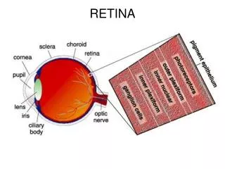
Rhodopsin= retinal (lipid) + opsin Different opsins=different photoreceptors (rods vs. cones, etc.) Vitamin A is a source of retinal Why are photoreceptors located where they are? Show animation.
RODS VERSUS CONES: approx 130,000 in eye • RODS: monochromatic, most sensitive to low light, on periphery, poor acuity (detail) • CONES: color, sensitive to mod/high light, concentrated in fovea, good acuity (why? We will discuss later). Make up only 3-5 % of the photoreceptors
Sensory transduction • Review: Amplifies (100k, 5 photon sensitive) • Note that light closes the sodium channels which causes a hyperpolarization. • This hyperpolarization causes a decrease in glutamate neurotransmitter release to a bipolar cell (more on this later) • Unique to the nervous system. So in darkness, the photoreceptors are depolarized. Evolution significance?
DARK/LIGHT ADAPTATION • To prepare for another photon, all trans retinal leaves the cone/rod, is metabolized back to 11-cis form and returned to the rod/cone. • How long does this take? • Rods:20-30 minutes. Respond to slow changes • Cones:2-3 minutes. Respond to more rapid changes.
What happens when we enter a dark movie theatre from outside in the sun? 99% of the retinal had been converted to the all trans and fallen off of the opsin There would be no signal until enough 11-cis production has occurred. Cones first (2-3 minutes) then rods(20-30 minutes) Pupil dilation
What happens when we walk out again from the movie theatre? Since we have been in the dark, we have regenerated all of the retinal into the 11-cis form. We walk out into the sun and every cell is ready to fire (blinding) until a short time later when the 11-cis is mostly converted to all trans and is used up (adjustment)
Light actually tells a photoreceptor to stop releasing glutamate. The glutamate then sends a signal to a bipolar nerve cell, which in turn, can also release glutamate if activated. This bipolar nerve cell can be an “on cell” which fires when light is shown. In order to do this, it therefore has to have a glutamate receptor that inhibits an action potential. An “off” bipolar nerve ganglion has a glutamate receptor that opens sodium channels and allows an action potential. review
Receptive fields • Think of the retina as a “screen” • Center surround organization • Our vision depends on viewing contrasts between objects and backgrounds • Rod receptive field larger than cones • “on”/”off” center surround organization • Concept of lateral inhibition, and rebound effect. Draw picture on board and show animation. Can explain after images and optical illusions.
Let us assume an “on” center and “off” surround field. In other words, the center of the receptive field has “on” ganglion cells (fire when photoreceptors receive light), while the surrounding ganglion cells are “off” when light is shown on them. Note that there are other fields where the opposite is occuring. Field A is more activated than field D. Field B is more activated than field C. When assuming an on center off surround field.
The surrounding “off” surround ganglion cells are inhibiting the “on” center ganglion cells in A more than B, creating a gray (less contrast image) spot When you look directly inbetween the grids, the spot goes away because receptive fields get smaller when you look directly at them.
Light contrast: to understand you must know the difference between intensity and lightness. Both are indicators of light reflecting off an object but…. intensity=physical and constant level lightness=subjective and perceptual. Can change based on differences of intensity between images. Each strip differs in intensity, but notice within each strip, lightness differs. In B the second light will inhibit the strength of the “on” ganglion in the center.
Field A detects the least amount of light, while field B detects the most amount of light. Fields C and D receive light from both the darker and lighter stripes, which creates antagonism. With field C, although its center detects the same intensity of light as field A (because they are located within the same stripe), part of its surround detects brighter light from the lighter stripe. As a result, the surround of field C creates more antagonism than the surround of field A, which decreases the activity of field C making the area around it appear darker in lightness. With field D, its center detects the same intensity of light as field B, but part of the surround is detecting the reduced light from the darker stripe. This creates less antagonism resulting in a lighter field.
Color vision • Do not confuse mixing colors of light and colors of pigment like paint • Pigments: yellow + blue = green. Mix all colors and you get black (subtractively). The more pigments you add the more wavelengths will be absorbed and the less reflected wavelengths that will reach the eye. • Light: yellow + blue = white (NOT GREEN) Red + green = yellow. The more lights you add, the closer you get to white light (additive). Newton and prisms. • The uniqueness of yellow.
Chemistry review • Visible light is a part of the electromagnetic energy spectrum • Photons of light are energy, and they travel in waves • Wavelengths are measured in nanometers (nm) • Visible light is only between 400nm and 750nm • Visible light spectrum ranges from Purple, Blue Green, Yellow, Orange, to Red • Below 400 is called Ultraviolet (Can’t See) • Above 750 is called Infrared (Can’t See) • Newton: “The Rays to speak properly are not coloured. In them there is nothing else than a certain Power and Disposition to stir up a Sensation of this or that Colour”. Wavelengths are not colored, color is created by our perceptual system in response to these wavelengths
Young & Helmholtz’s Trichromatic Theory of Color • You can combine three primary colors of light to reproduce all colors of the spectrum. Two colors are too few. • Therefore we have only 3 kinds of receptors to detect an infinite # of colors • Blue cones: have a unique opsin that maximally responds to 420 nm wavelength • Green cones: 530 nm • Red cones: 559 nm • No yellow cones! • Any single wavelength of visible light will stimulate a unique ratio of these 3 cones—allowing for an infinite # of colors (shades of colors)
Color wheels, spectrums etc. • We will review demonstrations • Again, remember that our perception of color depends on the ratio of excitation in the three different cone classes. Two different wavelength lights can evoke the same response (color perception) as long as the ratio of excitation is the same. These are called metamers • Max absorbency of photoreceptors is 555nm for us and old world apes. Evolution significance? • People can distinguish about 2 million different colors. 200 colors X 500 steps in intensity X 20 steps in saturation. Saturation=amount of whiteness in a color. Red is more saturated than pink
Opponency theory • Name a color that is yellowish red. How about reddish blue. How about bluish green. How about reddish green? How about bluish yellow? • In lateral geniculate nucleus (lgn) there are color-opponent neuron cells. This is similar to on/off center surround receptive fields. • Demonstration will explain
Color blindness • Most common types are protanopia and deuteranopia (95% of all color blindness) • See the world in shades of yellow and blue. Both red and green look yellowish. • Acuity is normal. • 7% of men are affected but only 0.4% of women affected. • What does this imply? What could be happening here?
Sex linked. The gene that codes for red cones and green cones must reside on the X chromosome. Since normal acuity, the red or green cones are actually being formed, but are not responding properly to the different wavelengths. (remember that color vision is dependent on “ratio” of excitation) Therefore, protanopes have their red cones filled up with green cone opsin. Deuteranopes have their green cones filled up with red opsin. If you don’t have different opsin molecules to respond to red and green wavelengths, then you cannot distinguish between red and green. Remember that yellow is perceived when red and green cones respond equally, which they would if they had the same opsin molecule.
More things to consider…. • The primates of South America, which broke from the continent of Africa about 40 million years ago, possess only a single functional copy of a red or green gene, much like color-blind men. Old World primates—the monkeys and apes of Africa and the ancestors of humans have two different opsin receptors for red and green like normal men. Researchers have sequenced human red and green opsin genes and discovered that they differ by only two percent. • Can you suggest an explanation for this? What might have happened to our ancestral genome in old world apes that led us to normally see red and green? What is the evolutionary significance of this?
A primordial red-green gene must have duplicated and then diverged slightly in sequence, leading to separate receptors of the red and green type. Being able to distinguish red berries in a green canopy might be advantageous. Curiously, researchers have also found that when they sequenced DNA in this region from some men who have normal color vision, they found that lying head to tail along their X chromosome were not just the two genes for the red and green receptors, but also an extra copy of the green receptor gene. This explains the why color blindness is a relatively common genetic disorder. What could be happening here? Hint, recall our studies with sry gene and recombination.
One X chromosome may receive an extra green receptor gene. This does no harm. But then the other chromosome with which it is exchanging bits of genetic information is left with only a red receptor gene. The man who inherits this slightly truncated chromosome will be color-blind, without the genetic information needed to make a green receptor.
Remember that more than 95 percent of all variations in human color vision involve the red and green receptors in men's eyes. It is very rare for anyone—male or female—to be "blind" to the blue end of the spectrum. Why do you think that blue end blindness is so rare? Do you think that it affects more men, or women?
The gene coding for the blue receptor lies on chromosome 7, which is shared equally by men and women, and this gene does not have any neighbor whose DNA sequence is similar. Blue color blindness is caused by a simple mutation in this gene.
Sacrifices peripheral vision(panorama) Total cross over gives panorama vision only We have approx. 50% crossover. For instance the left visual cortex receives optic nerve input from the left temporal region and the right nasal region. Parallax: Two slightly different images from each eye come together (fusion) in the brain creating a 3-D sensation (depth perception). This fusion process is a learned response and develops during infancy. If this didn’t happen we would have “double” vision. Convergence of eyes occur to help overcome double images.(one finger test). Lens focus is also important. Stereopsis is strongest with near objects. Why?(prove it to yourself) 3D vision depends on saccadic eye movements. It is optimal with images on the fovea. You can still have monocular depth perception. How? (movement stereo, etc.) 3D movies Binocular vision