Download
1 / 14
191 likes | 853 Vues
Enzyme Inhibition Test --- Inhibition of Succinate Dehydrogenase by Oxalate. PURPOSE. To understand the principles of enzyme inhibition To understand how to detect enzyme inhibition To understand the clinical significance of enzyme inhibition. Competitive i nhibition.

E N D
Enzyme Inhibition Test ---Inhibition of Succinate Dehydrogenase by Oxalate
PURPOSE • To understand the principles of enzyme inhibition • To understand how to detect enzyme inhibition • To understand the clinical significance of enzyme inhibition
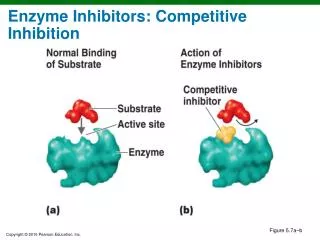
Competitive inhibition • Competitive inhibitors usually resemble the substrate • S and I “compete” for the enzyme active site • The relative concentration of substrate and inhibitor and their respective affinity with the enzyme determines thedegree of competitive inhibition. • The effect of a competitive inhibitor can be overcome with high concentrations of the substrate.
Clinical significance of enzyme inhibition • The usefulness of the most important pharmaceutical agents, antimetabolites, is based on the concept of competitive enzyme inhibition. • The antimetabolites are structural analogues of normal biochemical compounds. • As competitive inhibitors they compete with the naturally substrate for the active site of enzyme and block the formation of undesirable metabolic products in the body.
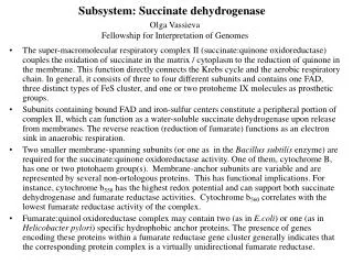
Succinate Dehydrogenase FAD FADH2 Succinate Fumarate • This enzyme coupled with the coenzyme FAD is bound to the inner membrane of the mitochondria. Succinate Dehydrogenase (SDH)
MBH2 (colorless) FAD succinate dehydrogenase MB (blue) FADH2 Fumarate • The enzymatic activity of succinate dehydrogenase can be measured by monitoring the reduction of an artificial electron acceptor, methylene blue (MB). • The oxidized form of the dye (MB) is blue and the reduced form (MBH2) is colorless.
FAD FADH2 COO COO Oxalate • In the reaction succinate → fumarate,oxalate (or malonate) has a structure similar to succinate and competes with it for the active site on succinate dehydrogenase. Competitive inhibition
Reagents and Materials 1. Phosphate buffer (pH 7.4) or 0.9% NaCl (NS,normal saline) 2. 0.25% Succinate solution 3. 0.5% Oxalate solution 4. Methylene blue (MB) 5. Mitochondria preparation: SDH is bound to the inner membrane of the mitochondria. Rat liver is an ideal source for functional intact mitochondria. We use rats for our studies.
PROCEDURE A.Preparation of Mitochondria from Rat Liver (1) Cervical dislocation • Press the head of the rat with the left thumb and forefingers. Grasp the base of the tail with the right thumb and forefingers and with a quick motion pull away and slightly upward from the skull. • This should cause a separation of the spinal cord from the brain.
(2) Preparation of Mitochondria • A medial incision (vertical, up the middle of the anima) from groin to sternum by scissors and tweezers, first separating the skin then the muscle and peritoneum, reveals the liver, which is dark brown and large. • Drop the liver in the mortar, cut it into pieces, grind by the pestle for homogenizing. Then add phosphate buffer 7.0ml (in batches, maybe 2+2+3ml) and grind after adding phosphate buffer each time. • Transfer the homogenate to a Centrifugal tube, centrifuge at 3,000 rpm (revolutions per minute) for 3 minutes. • Transfer the supernatant to another clean tube, which contains mitochondria . liver
Centrifugation • Centrifugation is a process that involves the use of the centrifugal force for the separation of mixtures. • More-dense components of the mixture migrate away from the axis of the centrifuge, while less-dense components of the mixture migrate towards the axis.
Centrifuge (<6,000rpm) rpm- revolutions per minute Microcentrifuge (<25,000rpm) Ultracentrifuge (>30,000rpm)
Notice for centrifugation • The liquid added to the centrifugal tubes should be even. Large difference may cause big shaking when in running state. • The tubes should be placed symmetrically by even number.
B. Label the test tubes according to following table and add the reagents • Mix the reagents in each tube and incubate at 37C (no shaking!). Look at disappearance of the blue color in each tube and record its turn and time, then analyze.