Download
1 / 20
200 likes | 326 Vues
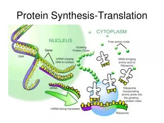
UV Radiation Regulates Mi-2 through Protein Translation and Stability. Craig J Burd, H. Karimi Kinyamu, Frederick W. Miller, and Trevor K Archer (2008). Melanie Cobb and Patrick English. Mi-2, The NuRD Complex. Known as the Nucleosome Remodeling Deacetylase Complex

E N D
UV Radiation Regulates Mi-2 through Protein Translation and Stability Craig J Burd, H. Karimi Kinyamu, Frederick W. Miller, and Trevor K Archer (2008) Melanie Cobb and Patrick English
Mi-2, The NuRD Complex Known as the Nucleosome Remodeling Deacetylase Complex 2 Isoforms, Mi-2a (CHD3) and Mi-2b (CHD4) Functions by Altering to Histone Tails (Histone Code) leading to Gene Repression by Compacting DNA Linked to Histone Deacetylase Activity Levels of this protein would naturally increase during the duplication of DNA NuRD Binding Domain of Mi-2b NuRD Function in Repression
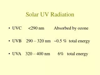
Dermatomyositis Disease State Overexpression of Mi-2 related to Autoimmune Disease Painful and/or itchy rash caused by inflammation of blood vessels under the skin and in the muscles Sudden or progressive weakness in muscles in neck, hip, back and shoulder muscles Difficulty swallowing (dysphagia), a feeling of choking Hardened lumps or sheets of calcium, called calcinosis, under the skin Changes in voice (dysphonia), especially hoarseness Image courtesy of the NIH.
Pg 3: Other NuRD complex proteins did NOT show similar upregulation in protein expression, EXCEPT MBD2, only a modest increase at higher UV doses. Cox-2 protein used as (+) control b/c known to increase w/ UV exposure. Surprise: only occurred at higher UV doses. Male/female Dif cell types
Flow Cytometry Cells are passed one at a time through Flow Cytometer and separated based on the cell’s reaction to light Propidium Iodide is Intercalating Agent, Fluorescence increased 30-40 nm when bound to Nucleic Acids Reasonable expectation that entering into S-Phase exposes DNA to binding by PI Propidium Iodide
PI Flow Cytometry No Change in PI levels seen until 8 hours after UV exposure However, Mi-2 levels in cell increase half an hour after UV exposure Demonstrates the change in Mi-2 expression is not due to cell cycle changes
Mi-2 labeled with Red Fluorescent Tag CPD Labelled with Green Fluorescent Tag Lack of Co-location after UV exposure indicates Mi-2 expression is not a result of DNA repair mechanisms Cyclobutane Pyramidine Dimer
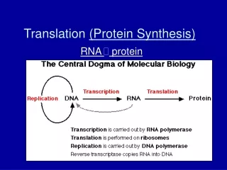
To Show that the Level of Mi-2 is Not Regulated Transciptionally • Cells were subjected to UV radiation • mRNA for each of the various NuRD proteins was collected at specific times • Quantification of mRNA • No significant changes in Mi-2α or Mi-2β were shown. • Strangely, other NuRD members changed. This is surprising because the level of protein did not increase after UV exposure, as shown in figure 1. They were confused.
To Show that the Level of Mi-2 is Not Regulated Transciptionally • To further ensure that the level of Mi-2 mRNA did NOT change after UV exposure, cells were treated with the RNA polymerase II inhibitor actinomycin D immediately after UV treatment. • These cells still showed an increase in Mi-2 protein level after UV exposure. • This verified that the change in protein level after UV treatment is translationally regulated, not transcriptionally regulated
UV Exposure Stabilized the Mi-2 Protein • Added the translational inhibitor cylcoheximide to keratinocytes after UV exposure • Collected cell lysates at various times afterwards • As shown above, there is a decrease in Mi-2 protein after 2 hours without UV treatment, but no decrease with UV treatment. • This indicates Mi-2 protein is stabilized with UV. • Also, since before 2 hours there is no difference in level of Mi-2 protein, it can be inferred that the increase in Mi-2 level observed after 30 minutes of UV exposure is NOT due to protein stabilization.
UV Exposure Stabilized the Mi-2 Protein • Wanted to further test the idea that the Mi-2 protein is stabilized after UV exposure • Treated keratinocytes with the proteosome inhibitor MG123. This pharmaceutically stabilized the protein by inhibiting its degradation. • In keratinocytes exposed to UV and MG123, Mi-2 is more stabilized than without UV exposure.
Is 5’ or 3’ UTR Responsible for Mi-2 Regulation? • The 3’ UTR and 5’UTR of Mi-2 were amplified with PCR and inserted into vector with the Luciferase reporter protein • Transfected keratinocytes (skin cells) with each vector • The cells are exposed to different experimental conditions, then the cell lysate harvested and tested for Luciferase activity.
Luciferase Firefly Luciferin • Luciferase is an bioluminescent enzyme that reacts with the substrate luciferin to emit light at a peak wavelength of 562 nm. • It is a good way to measure level of gene expression due to regulatory elements in genes (eg: UTR’s) without radioactivity. http://www.bdbiosciences.com/features/products/display_product.php?keyID=76
Is 5’ or 3’ UTR Responsible for Mi-2 Regulation? • All cells without UV had the same level of Luciferase activity, indicating the same level of Mi-2 expression in each cell, regardless of whether it has been transfected with a 5’ or 3’ UTR vector. • Two hours after exposure to UV, the Luciferase activity increased in cells transfected with 5’ regulatory region. • This indicates that the 5’ UTR region on the Mi-2 mRNA may be responsible for the increase level of translation of Mi-2 after UV exposure.
Is 5’ or 3’ UTR Responsible for Mi-2 Regulation? • To ensure that the increase in Luciferase activity is NOT due to an increase in translational activity, mRNA from the cells transfected with Lucerferase 3’ UTR, 5’ UTR, or no UTR was collected. • No substantial difference in mRNA levels with or without UV.
To Verify the Mechanism above Does Increase Mi-2 After UV • Cells grown in Methionine Free environment • Exposed to UV • 35S Methionine added • Immunoprecipitated and viewed by auto-radiography • Indeed, more Mi-2 in UV treated cells
Possible Explanation? • Increased Mi-2 expression from UV may provide the antigen presentation required for autoantibodies to form. • This would suggest that autoimmunity in many forms of myositis may be due to altered antigen presentation, rather than defective selectivity as in other autoimmune diseases. • From this study, we know that ½ hour after UV exposure, Mi-2 levels increase and are maintained for 16 hours. • This is due to translational regulation mediated by the 5’ UTR and protein stabilization in response to UV. This is not a result of an accumulation of S-phase cells. • Mi-2 does not co-localize with damaged DNA, and is not known to help repair damage.
• DM patients have photosensitive sensitive rashes, and tend to develop Mi-2 antibodies. UV exposure increases likelihood of DM and Mi-2 antibody development. • Mi-2, part of the NuRD complex, is increased and maintained after UV. However, other NuRD members are not. • It’s possible that Mi-2 is the limiting protein in complex Or that Mi-2 also acts independently of NuRD. • Mi-2 has been shown to be a transcriptional regulator.