Download
1 / 90
1.01k likes | 1.81k Vues
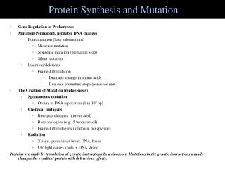
Translation: Protein Synthesis. Outline. Protein synthesis machinery: mRNA tRNA Aminoacyl-tRNA synthetases Ribosome Genetic code: Cracking the genetic code Codon usage Translation reaction: Initiation Elongation Termination

E N D
Outline • Protein synthesis machinery: • mRNA • tRNA • Aminoacyl-tRNA synthetases • Ribosome • Genetic code: • Cracking the genetic code • Codon usage • Translation reaction: • Initiation • Elongation • Termination • Difference between eukaryotes and prokaryotes in translation • Protein sorting • Inhibition of protein synthesis by antibiotics
Translation: biosynthesis of proteins • Translation is among the most conserved mechanisms across all organisms • It is the most energetically costly for the cell. Protein synthesis consumes about 80% energy of the cell • 50,000 ribosomes per E. coli cell • Ribosomes are the most complex polymerases. Molecular weight of a ribosome in E. coli is 2.7 million Daltons • Two thirds of the ribosome is RNA; Ribosomes can been see under EM • Translation requires: • amino acids, • mRNAs, • tRNAs, • aminoacyl tRNA synthetases, • ribosomes
The three roles of RNA in protein synthesis. Messenger RNA (mRNA) is translated into protein by the joint action of transfer RNA (tRNA) and the ribosome, which is composed of numerous proteins and two major ribosomal RNA (rRNA) molecules.
Three roles of RNA in translation • Messenger RNA (mRNA) carries the genetic information copied from DNA in the form of a series of three-base code "words," each of which specifies a particular amino acid. • Transfer RNA (tRNA) is the key to deciphering the code words in mRNA. Each type of amino acid has its own type of tRNA, which binds it and carries it to the growing end of a polypeptide chain if the next code word on mRNA calls for it. The correct tRNA with its attached amino acid is selected at each step because each specific tRNA molecule contains a three-base sequence that can base-pair with its complementary code word in the mRNA. • Ribosomal RNA (rRNA) associates with a set of proteins to form ribosomes. These complex structures, which physically move along an mRNA molecule, catalyze the assembly of amino acids into protein chains. They also bind tRNAs and various accessory molecules necessary for protein synthesis. Ribosomes are composed of a large and small subunit, each of which contains its own rRNA molecule or molecules.
mRNA • ORF (open reading frame):The protein coding region(s) of each mRNA is composed of a continous non-overlapping string of codons called an open reading frame (ORF). Each ORF specifies a single protein and starts with a start codon and ends with a stop codon. • In bacteria, start codons include 5’-AUG-3’ (the most common), 5’-GUG-3’ and 5’-UUG-3’. • In eukaryotes, the start codon is always 5’-AUG-3’. • The start codon not only specifies the first amino acids (aa) to be added, but it also defines the reading frame for subsequent codons. • Eukaryotic mRNAs mostly contain a single ORF (monocistronic mRNA), Prokaryotic mRNA usually contain multiple ORF (polycistronic mRNAs). • Prokaryotic mRNAs have a ribosome binding site (RBS) also called Shine-Dalgarno sequence. This sequence is complementary to the 16S rRNA of the small ribosomal subunit. • Eukaryotic mRNAs have a 5’ cap that binds ribosomes with the help of initiator proteins. Once bound, the ribosome scans the mRNA until it finds the first 5’-AUG-3’. Translation in eukaryotes is enhaced by the Kozak sequence: GCCACCAUGG and by the presence of a poly A tail.
tRNA • tRNAs are about 75-95 nucleotides long. The sequence from tRNA to tRNA varies, but there are common features to all tRNAs: a 3’ terminus 5’-CCA-3 (where the aa attaches), the presence of unusual bases and a common secondary structure. The secondary structure resembles a cloverleaf where there is an acceptor stem (5’-CCA-3’), three stem loops and the anticodon loop. • Attachment of aa to tRNA. tRNA molecules are charged when they have an aa attached. • Adenylylation: an amino acid reacts with ATP and becomes adenylylated (AMP-aa). A pyrophaosphate is released. This reaction is catalyzed by the aa specific tRNA synthetase. • tRNA charging: The adenylylated aa which remains tightly bound to the synthetase reacts with the tRNA. • The energy released when the bond is broken helps drive the formation of the peptide bond that links aa to each other.
Structure of tRNAs. (a) The primary structure of yeast alanine tRNA (tRNAAla), the first such sequence determined. This molecule is synthesized from the nucleotides A, C, G, and U, but some of the nucleotides, shown in red, are modified after synthesis: D = dihydrouridine, I = inosine, T = thymine, Y = pseudouridine, and m = methyl group. Although the exact sequence varies among tRNAs, they all fold into four base-paired stems and three loops. The partially unfolded molecule is commonly depicted as a cloverleaf. Dihydrouridine is nearly always present in the D loop; likewise, thymidylate, pseudouridylate, cytidylate, and guanylate are almost always present in the TYCG loop. The triplet at the tip of the anticodon loop base-pairs with the corresponding codon in mRNA. Attachment of an amino acid to the acceptor arm yields an aminoacyl-tRNA. (b) Computergenerated three-dimensional model of the generalized backbone of all tRNAs. Note the L shape of the molecule.
Aminoacyl-tRNA synthetases • Aminoacyl-tRNA synthetases from all organisms belong to one of two classes depending on the amino acid they are responsible for. Class I enzymes are generally (though not always) monomeric, and attach the carboxyl of their target amino acid to the 2' OH of adenosine 76 in the tRNA molecule. Class II enzymes are generally dimeric or tetrameric, and attach their amino acid to the 3' OH of their tRNA, except for phenylalaninyl-tRNA synthetase which uses the 2' OH. • Each of the 20 aa is attached to the appropriate tRNA by a single dedicated tRNA synthetase. The same tRNA synthetase is responsible for charging all tRNAs for a particular aa (for example when that aa has more than one codon). Thus most organisms have 20 different tRNA synthetases. • The acceptor stem and the anticodon loop of the tRNA specify to the tRNA synthetase which aa is to be added to the tRNA.
tRNA charging by aminoacyl-tRNA synthetase in two steps (proofreading) (proofreading) The amino acid is attached to the 3’OH of the ribose of the 3’ terminal A (in the CCA)
Aminoacyl-tRNA synthesis Amino acid + ATP + tRNA + H2O Aminoacyl-tRNA + AMP + 2Pi Amino acids are coupled to tRNAs through ester linkages to either the 2 - or the 3 -hydroxyl group of the 3 -adenosine residue. A linkage to the 3 -hydroxyl group is shown.
Ribosome • The ribosome contains ribosomal RNAs and proteins. The ribosome directs protein synthesis. • The ribosome consists of a large and small subunits. The large subunit contains the peptidyl transferase center (catalyses the formation of the peptide bond), and the factor bidnign center. The small subunit contains the decoding center. • In bacteria, the small subunit is 30S and the large subunit is 50S. The whole ribosome is 70S. In eukaryotes, the small subunit is 40S, the large subunit is 60S and the whole ribosome is 80S. • An mRNA bearing multiple ribosomes is known as a polyribosome or polysome. A single ribosome is in contact with about 30 nucleotides, but the large size of the ribosome allows a density of 1 ribosome for every 80 nucleotides of mRNA. The ability of multiple ribosomes to function on a single mRNA explain the relatively limited abundance of mRNA in the cell (about 1-5% of total RNA). • The ribosome contains 3 tRNA binding sites: The A site is the binding site for the aminoacylated tRNA; the P site is the binding site for the peptidyl-tRNA; the E site (for Exit) is the binding site for the uncharged tRNA. • The mRNA enters and exits the decoding center through 2 narrow channels in the small subunit.
The general structure of ribosomes in prokaryotes and eukaryotes. In all cells, each ribosome consists of a large and a small subunit. The two subunits contain rRNAs of different lengths, as well as a different set of proteins. All ribosomes contain two major rRNA molecules (dark red) 23S and 16S rRNA in bacteria, 28S and 18S rRNA in eukaryotes and one or two small RNAs (light red). The proteins are named L1, L2, etc., and S1, S2, etc., depending on whether they are found in the large or the small subunit
Ribosomes are the molecular machines that coordinate the interplay of charged tRNAs, mRNA, and proteins that leads to protein synthesis • RNA constitutes nearly two-thirds of the mass of the ribosomes. Ribosomal RNAs (5S, 16S, and 23S rRNA) play a central role in protein synthesis • Proteins are synthesized in the amino-to-carboxyl direction • Messenger RNA Is translated in the 5’-to-3’ direction • In prokaryotes, the start signal Is AUG (or GUG) preceded by several bases that pair with 16S rRNA • Ribosomes have three tRNA-binding sites that bridge the 30S and 50S subunits • The growing polypeptide chain is transferred between tRNAs on peptide-bond formation site of the large subunit • Only the codon-anticodon interactions determine the amino acid that is incorporated
The H. marismortui large ribosomal subunit. The rRNA (in gray) forms the core. The ribosomal proteins (yellow) are mostly on the surface. Two thirds of the ribosomal mass is rRNA. In this view, the surface of the subunit that interacts with the small subunit faces the reader. RNA is shown in gray. The backbones of the proteins visible are rendered in gold. The particle is approximately 250 Å across.
28S rRNA 18S rRNA Total RNA isolated from a human cell line, HeLa cells Total RNA from HeLa cells was prepared using different methods as shown in lanes 1, 2 and 3. RNA Marker (M) used ranged from 0.2 bp - 10 kb.
Breaking the entire genetic code by use of chemically synthesized trinucleotides. 20 ribosome-free bacterial extracts were prepared each containing all possible aminoacyl-tRNAs (tRNAs with an amino acid attached). In each sample, a different amino acid was radioactively labeled (green); the other 19 amino acids were bound to tRNAs but were unlabeled. Aminoacyl-tRNAs and trinucleotides passed through a nitrocellulose filter (left), but ribosomes were retained by the filter (center) and would bind trinucleotides and their cognate tRNAs (right). Each possible trinucleotide was tested separately for its ability to attract a specific tRNA by adding it with ribosomes to samples from each of the 20 aminoacyl-tRNA mixtures. The sample was then filtered. If the added trinucleotide caused the radiolabeled aminoacyl-tRNA to bind to the ribosome, then radioactivity would be detected on the filter (a positive test); otherwise, the label would pass through the filter (a negative test). By synthesizing and testing all possible trinucleotides, the researchers were able to match all 20 amino acids with one or more codons (e.g., phenylalanine with UUU as shown here).
Assigning codons using synthetic mRNAs containing a single ribonucleotide. Addition of such a synthetic mRNA to a bacterial extract that contained all the components necessary for protein synthesis except mRNA resulted in synthesis of polypeptides composed of a single type of amino acid as indicated. Poly-G did not work for this experiment. [See M. W. Nirenberg and J. H. Matthei, 1961, Proc. Nat'l. Acad. Sci. USA47:1588.]
Example of how the genetic code -- an overlapping, commaless triplet code -- can be read in two different frames. If translation of the mRNA sequence shown begins at two different upstream start sites (not shown), then two overlapping reading frames are possible; in this case, the codons are shifted one base to the right in the lower frame. As a result, different amino acids are encoded by the same nucleotide sequence. Many instances of such overlaps have been discovered in viral and cellular genes of prokaryotes and eukaryotes. It is theoretically possible for the mRNA to have a third reading frame.
Assigning codons using mixed polynucleotides (a) When a synthetic mRNA with alternating A and C residues was added to a protein-synthesizing bacterial extract, the resulting polypeptide contained alternating threonine and histidine residues. This finding is compatible with the two alternative codon assignments shown. (Note that alternating residues yield the same sequence of triplets regardless of which reading frame is chosen.) (b) To determine which codon assignment shown in (a) is correct, a second mRNA consisting of AAC repeats was tested. This mRNA, which can be read in three frames, yielded the three types of polypeptides shown. Since only the ACA codon was common to both experiments, it must encode threonine; thus CAC must encode histidine in (a). The assignments AAC = asparagine (Asn) and CAA = glutamine (Gln) were derived from additional experiments.
Francis Crick (center) with Gobind Khorana and Marianne Grunberg-Manago. Khorana unraveled much of the genetic code after Nirenberg’s initial breakthrough, which was based on Grunberg-Manago’s pioneering research. Gobind Khorana showed that tri- and tetra-nucleotides could be polymerized into polymers with repeating sequences that could be used in cell-free in vitro translation assays. Photo was taken at the 1966 Symposium on protein synthesis at Cold Spring Harbor Laboratory.
The codon usage trend is highly correlated to the accepting tRNA population of individual organisms;Codon usage may regulate gene expression at the translation level;A list of codon usage of different organisms can be found at the website: http://www.kazusa.or.jp/codon/
Ribosome is the assembly line for protein production The ribosome is a factory for the manufacture of polypeptides. Amino acids are carried into the ribosome, one at a time, connected to transfer RNA molecules (blue). Each amino acid is joined to the growing polypeptide chain, which detaches from the ribosome only once it is completed. This assembly line approach allows even very long polypeptide chains to be assembled rapidly and with impressive accuracy.
Initiation of translation in eukaryotes The translation always begins with a Met. When a ribosome dissociates at the termination of translation, the 40S and 60S subunits associate with initiation factors eIF3 and eIF6, forming complexes that can initiate another round of translation. Steps 1&2: sequential addition of the indicated components to the 40S subunit-eIF3 complex forms the initiation complex. Step 3: the initiation complex scans the mRNA until the start codon AUG is found. Step 4: association of the 60S subunit forms the 80S ribosome ready to translate the mRNA. Two initiation factors, eIF2 and eIF5, are GTP-binding proteins, those bound GTP is hydrolyzed during translation initiation.
Elongation during translation in eukaryotes Once the 80S ribosome with Met-tRNAiMet in the ribosome P site is assembled, a complex bearing the second amino acid (aa2) coded by the mRNA binds to the A site (step 1). Following a conformational change in the ribosome induced by hydrolysis of the GTP in EF1a.GTP (step 2), the large rRNA catalyzes peptide bond formation between Meti and aa2 (step 3). Hydrolysis of GTP in EF2.GTP causes conformational change in the ribosome that results in its translocation one codon along the mRNA and shifts the free tRNAiMet to the E site and the tRNA with the bound peptide to the P site (step 4). The cycle can begin again with binding of another complex bearing aa3 to the now open A site. The free tRNA at the E site is ejected during the step 2 in the next cycle. This cycle can go on until the ribosome reaches the stop codon on the mRNA.
Transfer RNA-Binding Sites. (A) Three tRNA-binding sites are present on the 70S ribosome. They are called the A (for aminoacyl), P (for peptidyl), and E (for exit) sites. Each tRNA molecule contacts both the 30S and the 50S subunit. (B) The tRNA molecules in sites A and P are base paired with mRNA.
Termination of translation in eukaryotes When a ribosome bearing a nascent protein chain reaches a stop codon (UAA, UAG or UGA), release factor eRF1 together with eRF3.GTP enter the ribosome complex. Hydrolysis of the bound GTP is accompanied by cleavage of the peptide chain from the tRNA in the P site and release of the tRNAs and two ribosomal subunits.
Model of protein synthesis on circular polysomes and recycling of ribosomal subunits. Multiple individual ribosomes can simultaneously translate a eukaryotic mRNA. The circular mRNA is stabilized by interactions between proteins bound at the 3’ and 5’ ends. When a ribosome completes translation and dissociates from the 3’ end, the separated subunits can rapidly find the nearby 5’ cap and initiate another round of synthesis.
Initiation of translation in prokaryotes • A charged tRNA enters the P site directly. This requires an initiator tRNA. In prokaryotes, the initiator tRNA is N-formyl methionine-tRNAi or fMet-tRNAiMet. • Three initiator factors (IF) are required: • IF3 binds to the small subunit and blocks it from re-associating with the large subunit, or from binding charged tRNAs. • IF1 prevents tRNAs from binding to portions of the small subunit that will become part of the A site. • IF2 is a GTPase that interacts with the small subunit, IF1 and fMet-tRNAiMet. • With all 3 IFs bound, the small subunit is prepared to bind to the mRNA and the initiator tRNA (fMet-tRNAiMet). • The last step of initiation involves the association of the large subunit to create the 70S initiation complex. When the start codon and fMet-tRNAiMet base pair, the small subunit changes conformation and IF3 is released. The large subunit binds and stimulates GTPase activity of IF2-GTP. IF2-GDP has low affinity for the ribosome and fMet-tRNAiMet and is released along with IF1.
Formylation of methionyl-tRNA in bacteria Initiator tRNA (tRNAf) is first charged with methionine, and then a formyl group is transferred to the methionyl- tRNAf from N10-formyltetrahydrofolate.
Translation initiation in prokaryotes Prokaryotic protein synthesis differs from eukaryotic protein synthesis primarily in translation initiation: Ribosomes: Eukaryotic ribosomes are larger, 80S (60S and 40S subunits) with a mass of 4200 kd. Prokaryotic ribosomes are smaller, 70S (50S and 30S) with a mass of 2700 kd. Initiator tRNA: In eukaryotes, the initiating amino acid is methionine. In prokaryotes it is N-formylmethionine. Initiation: The initiation codon in eukaryotes is always AUG, the AUG nearest the 5’ end of mRNA is usually selected as the start site. Eukaryotic mRNA encodes only one protein hence it has only one start site. In prokaryotes, the initiation codon usually is AUG, or GUG with less frequency. A prokaryotic mRNA can encode multiple proteins hence it can have several translation start sites directed by several Shine-Dalgarno sequences.
Translation initiation in prokaryotes and eukaryotes Prokaryotic mRNA initiation occurs at AUG codons with properly spaced Shine-Delgarno sequence 5’ AUG SD AUG AUG SD AUG 3’ initiation codon with Shine-Delgarno site internal Met codon does not have Shine-Delgarno site initiation codon with Shine-Delgarno site Shine-Delgarno (SD) site consists of 3-9 contiguous bases in the mRNA that base pair with the 3’ end of 16S rRNA and is located optimally 5 nt upstream of the initiator codon Eukaryotic mRNA initiation occurs ~90% of the time at the first AUG codon 5’ cap AUG AUG AUG 3’ first AUG codon downstream of the 5’ cap internal Met codon
Translation initiation sites in prokaryotes: the Shine-Dalgarno sequence The Shine-Dalgarno Sequence (AGGAGGU) is the signal for initiation of protein biosynthesis in bacterial mRNAs. It is located 5' of the first coding AUG, and consists primarily, but not exclusively, of purines. The complementary sequence (ACCUCCU), rich in pyrimidines, is called the Anti-Shine-Dalgarno sequence and is located at the 3' end of the 16S rRNA in the ribosome. Mutations in the Shine-Dalgarno Sequence can reduce translation.
Complementary base pairing occurs between the 3' end of 16S rRNA and the region near the 5' end of an mRNA
GTP-driven translocation in prokaryotes by elongation factor G (EF-G) The cycle of elongation begins with peptidyl-tRNA in the P site. An aminoacyl-tRNA binds in the A site. With both sites occupied, a new peptide bond is formed. The tRNAs and the mRNA are translocated through the action of elongation factor G (EF-G), which moves the deacylated tRNA to the E site. Once there, it is free to dissociate to complete the cycle.
Differences in elongation and termination between prokaryotes and eukaryotes • Eukaryotic elongation factors EF1a and EF1bg are the counterparts of prokaryotic EF-Tu and EF-Ts. The GTP form of EF1a delivers aminoacyl-tRNA to the A site of the ribosome, and EF1bg catalyzes the exchange of GTP for bound GDP. • Eukaryotic EF2 mediates GTP-driven translocation in much the same way as does prokaryotic EF-G. • Termination in eukaryotes is carried out by a single release factor, eRF1, compared with two in prokaryotes. • eIF3, like its prokaryotic counterpart IF3, prevents the re-association of ribosomal subunits in the absence of an initiation complex.
DNA mRNA Protein Ribosome binding site (RBS) or Shine-Dalgarno sequence directs the synthesis of multiple proteins from one mRNA