Chapter 13 Hemorrhage and Shock
800 likes | 1.05k Vues
Chapter 13 Hemorrhage and Shock. Objectives (1 of 8). 1.6.35 Describe the anatomy of the skin, bones, vessels, and subcutaneous tissue as it relates to hemorrhage control. 1.6.36 Discuss the benefits and complications of hemorrhage control by the following means: Direct pressure Tourniquets

Chapter 13 Hemorrhage and Shock
E N D
Presentation Transcript
Objectives (1 of 8) • 1.6.35 Describe the anatomy of the skin, bones, vessels, and subcutaneous tissue as it relates to hemorrhage control. • 1.6.36 Discuss the benefits and complications of hemorrhage control by the following means: • Direct pressure • Tourniquets • Hemostats • 1.6.40 Define shock.
Objectives (2 of 8) • 1.8.1 Define shock based on aerobic and anaerobic metabolism. • 1.8.2 Discuss the prevention of anaerobic metabolism. • 1.8.3 Discuss red blood cell oxygenation in the lungs based on alveolar O2 levels and transportation across the alveolar capillary wall. • 1.8.4 Discuss tissue oxygenation based on tissue perfusion and release of oxygen.
Objectives (3 of 8) • 1.8.5 Discuss the role played by respiration, inadequate ventilation in the management of shock. • 1.8.6 Describe perfusion and the mechanisms of improvement of cardiac output based on the strength and rate of contractions. • 1.8.7 Discuss the fluid component of the cardiovascular system and the relationship between the volume of the fluid and the size of the container.
Objectives (4 of 8) • 1.8.8 Discuss the systemic vascular resistance, the relationship of diastolic pressure to the SVR and the effect of diastolic pressure on coronary circulation. • 1.8.9 Discuss the container size in its relationship to the fluid volume and the effect on blood returning to the heart. • 1.8.21 Describe the mechanism of the body response to perfusion change. • 1.8.22 Identify the role of the baroreceptor.
Objectives (5 of 8) • 1.8.23 Describe how the actions of the baroreceptor affect blood pressure and perfusion. • 1.8.24 Describe compensated shock. • 1.8.25 Describe uncompensated shock, both cardiac and peripheral effects. • 1.8.26 Discuss the assessment of the patient’s perfusion status, based on physical observations within the primary survey, including pulse, skin, temperature, and capillary refill.
Objectives (6 of 8) • 1.8.27 Discuss the relationship of the neurological exam to assessment of hypoperfusion and oxygenation. • 1.8.28 Describe the information provided by the following in physical examination: pulse, blood pressure, diastolic pressure, systolic pressure, skin color, appearance, temperature, and respiration.
Objectives (7 of 8) • 1.8.29 Discuss management of a shocky patient. Include red cell oxygenation, tissue ischemic sensitivity, IV fluids, and the pneumatic antishock garment. • 1.8.30 Describe the beneficial and detrimental effects of the pneumatic antishock garment. • 1.8.31 Describe the indications and contraindications for the pneumatic antishock garment.
Objectives (8 of 8) • S1.8.35 Demonstrate in order of priority the steps of shock resuscitation. • S1.8.36 Demonstrate the use of the pneumatic antishock garment (PASG).
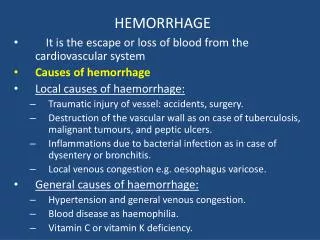
Bleeding (Hemorrhage) • Can be external and obvious or internal and hidden. • Causes weakness and eventually shock and death. • Most common cause of shock after trauma.
Shock • A state of collapse and failure of the cardiovascular system in which blood circulation slows and eventually ceases. • Can be fatal. • Accompanies events like heart attacks and automobile crashes.
Perfusion (1 of 2) • Circulation within tissues in adequate amounts to meet the cells’ needs for oxygen, nutrients, and waste removal. • Some tissues and organs need a constant supply of blood whereas others can survive on very little when at rest.
Perfusion (2 of 2) • The heart demands a constant supply of blood. • The brain and spinal cord can survive for 4 to 6 minutes without perfusion. • The kidneys may survive 45 minutes. • The skeletal muscles may last 2 hours.
Cardiovascular System (2 of 3) • Function • Circulates blood • Delivers oxygen and nutrients • Carries away waste
Cardiovascular System (3 of 3) • Components • Pump (heart) • Container (vessels) • Fluid (blood)
Blood Vessels • Arteries • Arterioles • Capillaries • Venules • Veins
Blood • Contains: • Red blood cells • White blood cells • Platelets • Plasma
Scene Safety • Follow BSI precautions. • Wear gloves and eye protection in all situations. • Avoid direct contact with body fluids. • Thorough hand washing between patients and after runs is important.
External Bleeding • Hemorrhage = bleeding • Body cannot tolerate greater than 20% blood loss. • Blood loss of 1 L can be dangerous in adults; in children, loss of 100-200 mL is serious.
Internal Bleeding • Internal bleeding may not be readily apparent. • Assess patient’s • Mechanism of injury • Nature of illness
Mechanism of Injury Can Indicate Internal Bleeding • When mechanism of injury suggests that severe forces affected the abdomen and/or the chest. • As a result of falls, blast injuries, and automobile or motorcycle crashes. • With penetrating injury, such as a knife or gunshot wound.
Nature of Illness Can Suggest Internal Bleeding • In the abdomen as a result of irritable bowel syndrome, an aneurysm, or a ruptured ectopic pregnancy. • Gastrointestinal problems may cause vomiting of blood or bloody diarrhea.
Signs and Symptoms of Internal Bleeding (1 of 2) • Ecchymosis: Bruising • Hematoma: Bleeding beneath the skin • Hematemesis: Blood in vomit • Melena: Black, tarry stool
Signs and Symptoms of Internal Bleeding (2 of 2) • Hemoptysis: Coughing up blood • Pain, tenderness, bruising, guarding, or swelling • Broken ribs, bruises over the lower chest, or rigid, distended abdomen
Significance of Bleeding • Body will not tolerate blood loss greater than 20% of blood volume. • More than 1 L of blood loss in an adult causes increased heart rate and decreased blood pressure. • Smaller amount of blood loss in infants and children causes significant effects. • Low blood volume results in inadequate perfusion and death.
Conditions With Possible Serious Bleeding • Significant mechanism of injury. • Poor general appearance of patient. • Assessment reveals signs of shock. • Significant amount of blood loss noted. • The blood loss is rapid. • You cannot control external bleeding.
Physiologic Response to Hemorrhage (1 of 2) • Arterial • Blood is bright red and spurts. • Venous • Blood is dark red and does not spurt. • Capillary • Blood oozes out and is controlled easily.
Blood Clotting • Bleeding normally stops within 10 minutes. • Some medications interfere with clotting. • Some injuries will be unable to clot. • Patients with hemophilia lack clotting factors.
Stage One Hemorrhage (1 of 2) • Up to 15% intravascular loss. • Compensated by constriction of vascular bed. • Blood pressure maintained. • Normal pulse pressure, respiratory rate, and renal output.
Stage One Hemorrhage (2 of 2) • Pallor of the skin. • Central venous pressure low to normal.
Stage Two Hemorrhage (1 of 2) • 15-25% intravascular loss. • Cardiac output cannot be maintained by arteriolar constriction. • Reflex tachycardia. • Increased respiratory rate. • Blood pressure maintained. • Catecholamines increase peripheral resistance.
Stage Two Hemorrhage (2 of 2) • Increased diastolic pressure. • Narrow pulse pressure. • Diaphoresis from sympathetic stimulation. • Renal output near normal.
Stage Three Hemorrhage • 25-35% intravascular loss • Classic signs of hypovolemic shock • Marked tachycardia • Marked tachypnea • Decreased systolic pressure • Decreased urine output • Alteration in mental status • Diaphoresis with cool, pale skin
Stage Four Hemorrhage • Loss greater than 35%. • Extreme tachycardia. • Pronounced tachypnea. • Significantly decreased systolic blood pressure. • Confusion and lethargy. • Skin is diaphoretic, cool, and extremely pale.
Assessment • Ensure an open and patent airway. • Have suction ready for bleeding of the mouth or facial areas. • Note the color of bleeding and try to determine its source. • Coffee-ground emesis is a sign of upper GI bleeding.
Controlling External Bleeding • Follow BSI precautions. • Ensure patient has an open airway and adequate breathing. • Provide oxygen if necessary. • Assess location and color of blood. • There are several methods to control bleeding.
Direct Pressure and Elevation • Direct pressure is the most common and effective way to control bleeding. • Apply pressure with gloved finger or hand. • Elevating a bleeding extremity often stops venous bleeding. • Use both direct pressure and elevation whenever possible. • Apply a pressure dressing.
Pressure Points • If bleeding continues, apply pressure on pressure point. • Pressure points are located where a blood vessel lies near a bone. • Be familiar with the location of pressure points.
Splints • Splints can help control bleeding associated with a fracture. • Air splints can be used to control bleeding of soft-tissue injuries.
Hemostats • Use on retracted vessels. • Apply to end of vessel.
Pneumatic Antishock Garment (PASG) • Stabilizes fractures of the pelvis and proximal femurs. • Controls significant internal bleeding. • Controls massive soft-tissue bleeding of the lower extremities.
PASG Contraindications • Pregnancy (do not inflate abdomen) • Pulmonary edema of cardiac origin • Acute heart failure • Penetrating chest injuries • Groin injuries • Major head injuries • Less than 30 minutes transport time
Application of PASG • Apply the garment so the top is below the lowest rib. • Enclose both legs and the abdomen. • Open the stopcocks. • Inflate with the foot pump. • Check patient’s vital signs.
Applying a Tourniquet • Fold a triangular bandage into 4” cravat. • Wrap the bandage. • Use a stick as a handle to twist and secure the tourniquet. • Write “TK” and time and place on patient.
Tourniquet Precautions • Place as close to injury as possible, but not over joint. • Never use narrow material. • Use wide padding under the tourniquet. • Never cover a tourniquet with a bandage. • Do not loosen the tourniquet once applied.
Bleeding From the Nose, Ears, and Mouth • Causes: • Skull fractures • Facial injuries • Sinusitis • High blood pressure • Coagulation disorders • Digital trauma