Shock and Hemorrhage
Shock and Hemorrhage . M. Alhashash MD, lecturer of general surgery, hepatobiliary & liver transplant surgery . malhashash@gmail.com 01093368234. Defining Shock. Shock is best defined as inadequate tissue perfusion. Can result from a variety of disease states and injuries.

Shock and Hemorrhage
E N D
Presentation Transcript
Shock and Hemorrhage M. Alhashash MD, lecturer of general surgery, hepatobiliary & liver transplant surgery. malhashash@gmail.com 01093368234
Defining Shock • Shock is best defined as inadequate tissue perfusion. • Can result from a variety of disease states and injuries. • Can affect the entire organism, or it can occur at a tissue or cellular level.
Defining Shock (2 of 2) • Shock is not adequately defined by: • Pulse rate • Blood pressure • Cardiac function • Hypovolemia • Loss of systemic vascular resistance
“Hypoperfusion can be present in the absence of significant hypotension.”
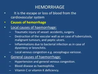
Definitions • Hemorrhage • Abnormal internal or external loss of blood • Homeostasis • Tendency of the body to maintain a steady and normal internal environment • Shock • INADEQUATE TISSUE PERFUSION • Transition between homeostasis and death
Definitions • O2 Delivery - volume of gaseous O2 delivered to the tissue/min. • O2 Consumption - volume of gaseous O2 which is actually used by the tissue/min. • O2 Demand - volume of O2 actually needed by the tissues to function in an aerobic manner Demand > consumption = anaerobic metabolism
Cardiovascular System Regulation to maintain volume and oxygen supply. • Neural • hormonal
Cardiovascular System Regulation Sympathetic Nervous System • Increase • Body activity • Heart rate • Strength of contractions • Vascular constriction • Bowel and digestive viscera • Decreased urine production • Bronchodilation • Increases skeletal muscle perfusion
BaroreceptorReflexes • High in the neck, each carotid artery divides into external and internal carotid arteries. • At the bifurcation, the wall of the artery is thin and contains many vine-like nerve endings. • The small portion of the artery is the carotid sinus. • Nerve endings in this area are sensitive to stretch or distortion. • Serve as pressure receptors or baroreceptors.
BaroreceptorReflexes • Similar area found in the arch of the aorta. • Serves as a second important baroreceptor • Large arteries, large veins, and the wall of the myocardium also contain less important baroreceptors. • Baroreceptor reflexes help maintain blood pressure by two negative feedback mechanisms: • By lowering blood pressure in response to increased arterial pressure • By increasing blood pressure in response to decreased arterial pressure
BaroreceptorReflexes • Normal blood pressure partially stretches the arterial walls so that baroreceptors produce a constant, low-frequency stimulation. • Impulses from the baroreceptors inhibit the vasoconstrictor center of the medulla and excite the vagal center when blood pressure increases. • Results in vasodilation in the peripheral circulatory system and a decrease in the heart rate and force of contraction. • Combined effect is a decrease in arterial pressure.
Baroreceptor Reflexes • Baroreceptorsadapt in 1 to 3 days to whatever pressure level they are exposed. Therefore, they do not change the average blood pressure on a long-term basis. This adaptation is common in people who have chronic hypertension.
BaroreceptorReflexes • When baroreceptor stimulation ceases due to a fall in arterial pressure, several cardiovascular responses are evoked: • Vagal stimulation is reduced and sympathetic response is increased. • The increase in sympathetic impulses results in increased peripheral resistance and an increase in heart rate and stroke volume. • Sympathetic discharges also produce generalized arteriolar vasoconstriction, which decreases the container size.
Chemoreceptor Reflexes • Chemoreceptors • Monitor level of CO2 in CSF • Monitor level of O2 in blood
Chemoreceptor Physiology • Low arterial pressure leads to hypoxemia and/or acidosis. • Hypoxemia/acidosis stimulate peripheral chemoreceptor cells within the carotid and aortic bodies. • These bodies have an abundant blood supply. • When oxygen or pH decreases, these cells stimulate the vasomotor center of the medulla. • The rate and depth of ventilation are also increased to help eliminate excess carbon dioxide and maintain acid-base balance.
CV System and Hormone Regulation • Catecholamines • Epinephrine • Norepinephrine
CV System and Hormone Regulation • Antidiuretic Hormone (ADH)(water & pressure regulation) • Release • Posterior pituitary • Drop in BP or increase in serum osmolarity • Action • Increase in peripheral vascular resistance • Increase water retention by kidneys • Decrease urine output • Splenic vasoconstriction • 200 mL of free blood to circulation
CV System and Hormone Regulation • Angiotensin II • Release • Primary chemical from kidneys • Lowered BP and decreased perfusion • Action • Converted from renin into angiotensin I • Modified in lungs to angiotensin II • 20-minute process • Potent systemic vasoconstrictor • 1-hour duration • Causes release of ADH, aldosterone, and epinephrine
CV System and Hormone • Aldosterone • Release • Adrenal cortex • Stimulated by angiotensin II • Action • Maintain kidney ion balance • Retention of sodium and water • Reduce insensible fluid loss
CV System and Hormone • Glucagon • Release • Alpha cells of pancreas • Triggered by epinephrine • Action • Causes liver and skeletal muscles to convert glycogen into glucose • Gluconeogenesis
Insulin Release Beta cells of pancreas Action Facilitates transport of glucose across cell membrane Erythropoietin Release Kidneys Hypoperfusion or hypoxia Action Increases production and maturation of RBCs in the bone marrow CV System and Hormone Regulation
Reabsorption of Tissue Fluids • Arterial hypotension, arteriolar constriction, and reduced venous pressure during hypovolemialower the blood pressure in the capillaries (hydrostatic pressure). • The decrease promotes reabsorption of interstitial fluid into the vascular compartment. • Considerable quantities of fluid may be drawn into the circulation during hemorrhage.
Reabsorption of Tissue Fluids • Approximately 0.25 mL/min/kg of body weight (1 L/hr in the adult male) can be autotransfused from the interstitial spaces after acute blood loss.
Blood • Blood Volume • Average adult male has a blood volume of 7% of total body weight. • Average adult female has a blood volume of 6.5% of body weight. • Normal adult blood volume is 4.5–5 L. • Remains fairly constant in the healthy body.
Blood Components • Erythrocyte: 45% • Hemoglobin • Hematocrit • Miscellaneous blood products: <1% • Platelets • Leukocytes • Monocytes, basophils, esonophils, neutrophils • Plasma: 54%
Signs of Organ Hypoperfusion • Mental Status Changes • Oliguria • Lactic Acidosis
Categories of Shock • HYPOVOLEMIC • CARDIOGENIC • DISTRIBUTIVE • OBSTRUCTIVE
Four Stages • Stage 1: Vasoconstriction • Stage 2: Capillary and venule opening • Stage 3: Disseminated intravascular coagulation • Stage 4: Multiple organ failure
Stage 1: Vasoconstriction (1 of 4) • < 15 % loss of blood volume. • Vasoconstriction begins as minimal perfusion to capillaries continues. • Oxygen and substrate delivery to the cells supplied by these capillaries decreases. • Anaerobic metabolism replaces aerobic metabolism.
Stage 1: Vasoconstriction (2 of 4) • Production of lactate and hydrogen ions increases. • The lining of the capillaries may begin to lose the ability to retain large molecular structures within its walls. • Protein-containing fluid leaks into the interstitial spaces (leaky capillary syndrome).
Stage 1: Vasoconstriction (3 of 4) • Sympathetic stimulation produces: • Pale, sweaty skin • Rapid, thready pulse • Elevated blood glucose levels • The release of epinephrine dilates coronary, cerebral, and skeletal muscle arterioles and constricts other arterioles. • Blood is shunted to the heart, brain, skeletal muscle, and capillary flow to the kidneys and abdominal viscera decreases.
If this stage of shock is not treated by prompt restoration of circulatory volume, shock progresses to the next stage. Stage 1: Vasoconstriction (4 of 4)
Stage 2: Capillary and Venule Opening (1 of 5) • Stage 2 occurs with a 15% to 25% decrease in intravascular blood volume. Heart rate, respiratory rate, and capillary refill are increased, and pulse pressure is decreased at this stage. Blood pressure may still be normal.
Stage 2: Capillary and Venule Opening (2 of 5) • As the syndrome continues, the precapillary sphincters relax with some expansion of the vascular space. • Postcapillary sphincters resist local effects and remain closed, causing blood to pool or stagnate in the capillary system, producing capillary engorgement.
Stage 2: Capillary and Venule Opening (3 of 5) • As increasing hypoxemia and acidosis lead to opening of additional venules and capillaries, the vascular space expands greatly. • Even normal blood volume may be inadequate to fill the container. • The capillary and venule capacity may become great enough to reduce the volume of available blood for the great veins and vena cava. • Resulting in decreased venous return and a fall in cardiac output.
Stage 2: Capillary and Venule Opening (4 of 5) • Low arterial blood pressure and many open capillaries result in stagnant capillary flow. • Sluggish blood flow and the reduced delivery of oxygen result in increased anaerobic metabolism and the production of lactic acid. • The respiratory system attempts to compensate for the acidosis by increasing ventilation to blow off carbon dioxide.
Stage 2: Capillary and Venule Opening (5 of 5) • As acidosis increases and pH falls, the RBCs may cluster together (rouleaux formation). • Halts perfusion • Affects nutritional flow and prevents removal of cellular metabolites • Clotting mechanisms are also affected, leading to hypercoagulability. • This stage of shock often progresses to the third stage if fluid resuscitation is inadequate or delayed, or if the shock state is complicated by trauma or sepsis.
Stage 3: Disseminated Intravascular Coagulation (DIC) (1 of 4) • Time of onset will depend on degree of shock, patient age, and pre-existing medical conditions. • Stage 3 occurs with 25% to 35% decrease in intravascular blood volume. At this stage, hypotension occurs. This stage of shock usually requires blood replacement.
Stage 3: Disseminated Intravascular Coagulation (DIC) (2 of 4) • Stage 3 is resistant to treatment (refractory shock), but is still reversible. • Blood begins to coagulate in the microcirculation, clogging capillaries. • Capillaries become occluded by clumps of RBCs. • Decreases capillary perfusion and prevents removal of metabolites • Distal tissue cells use anaerobic metabolism, and lactic acid production increases.
Stage 3: Disseminated Intravascular Coagulation (DIC) (3 of 4) • Lactic acid accumulates around the cell. • Cell membranes no longer have the energy needed to maintain homeostasis. • Water and sodium leak in, potassium leaks out, and the cells swell and die.
Stage 3: Disseminated Intravascular Coagulation (DIC) (4 of 4) • Pulmonary capillaries become permeable, leading to pulmonary edema. • Decreases the absorption of oxygen and results in possible alterations in carbon dioxide elimination • May lead to acute respiratory failure or adult respiratory distress syndrome (ARDS) • If shock and disseminated intravascular coagulation (DIC) continue, the patient progresses to multiple organ failure.
Stage 4: Multiple Organ Failure (1 of 2) • The amount of cellular necrosis required to produce organ failure varies with each organ and the underlying condition of the organ. • Usually hepatic failure occurs, followed by renal failure, and then heart failure. • If capillary occlusion persists for more than 1 to 2 hours, the cells nourished by that capillary undergo changes that rapidly become irreversible. • In this stage, blood pressure falls dramatically (to levels of 60 mmHg or less). • Cells can no longer use oxygen, and metabolism stops.
Stage 4: Multiple Organ Failure (2 of 2) • If a critical amount of the vital organ is damaged by cellular necrosis, the organ soon fails. • Failure of the liver is common and often presents early. • Capillary blockage may cause heart failure. • GI bleeding and sepsis may result from GI mucosal necrosis. • Pancreatic necrosis may lead to further clotting disorders and severe pancreatitis. • Pulmonary thrombosis may produce hemorrhage and fluid loss into the alveoli. • Leading to death from respiratory failure.
Goals of Shock Resuscitation • Restore blood pressure • Normalize systemic perfusion • Preserve organ function