Myocardial Ischemia
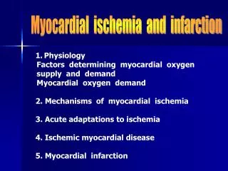
Myocardial Ischemia. Ischemia. Is when blood flow to the myocardium is insufficient to maintain the metabolic demand of the myocytes. Transmural Ischemia

Myocardial Ischemia
E N D
Presentation Transcript
Ischemia • Is when blood flow to the myocardium is insufficient to maintain the metabolic demand of the myocytes. Transmural Ischemia • The hallmark of acute transmural ischemia (across the heart wall from endocardium to epicardium ) is the elevation of the ST segment of the ECG. • This is visualized by the ST-segment being raised above the isoelectric baseline. • This is due to a higher (more positive) resting voltage of ischemic cells.
Which cause the ST-segment baseline to be more positive (an upwards deflection). • This change in the ST segment is mostly localized in the lead most directly overlying the ischemic myocardial area. • This may lead to unstable angina, and should be taken very seriously, as this is the condition that progresses to an acute MI. • As the ischemia becomes more extensive the ST segment elevation becomes more pronounced.
Sub-Endocardial Ischemia • Relative decreased flow in the subendocardialregions is a normal consequence of the squeezing of the myocardium, which compresses the blood supply to the endocardium during ventricular systole. • However, this can also be a pathological condition, expressed in stable-angina. This is a condition where the myocardial demands exceed the coronary artery blood supply .
This is represented on the ECG as ST-segment depression. • Theischemic cells become more positive in their resting voltage (due to channel leakage) and this makes the subendocardium more positive; since the endocardium is further away from the precordial leads than the more negative mycardium we see this as a depression .
After the ischemia has progressed to an infarct, and the tissue has scarred, the ECG will show • Q wave = Necrosis (significant Q’s only) • Significant Q wave is one millimeter (one small square) wide, which is .04 sec. in duration or more or is a Q wave >1/4the amplitude (or more) of the QRS complex. • Old infarcts: significant Q waves (like infarct damage) remain for a lifetime.
ST (segment) elevation = (acute) Injury) • Signifies an acute process, ST segment returns to baseline with time. • T wave inversion = Ischemia • Inverted T wave (of ischemia) is symmetrical (left half and right half are mirror images). Normally T wave is upright when QRS is upright, and vice versa. • Usually in the same leads that demonstrate signs of acute infarction (Q waves and ST elevation).
Infarction Location: • Lateral Q’s in lateral leads I and AVL (Circumflex Coronary Artery). • Anterior Q’s in V1, V2, V3, and V4 (Anterior Descending Coronary Artery) • Inferior (diaphragmatic) • Q’s in inferior leads II, III, and AVF (R. or L. Coronary Artery).