Download
1 / 20
200 likes | 552 Vues
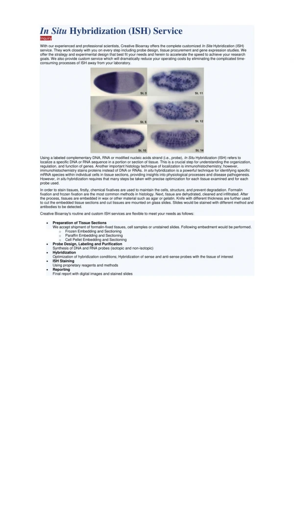
The effects of NAFLD on the liver sinusoidal endothelial cells. By Edward Harris Dept. of Biochemistry. Liver structure overview. Stellate cell. Space of Disse. Kupffer cell. Hepatocyte. Endothelial cell. Sasse et al. 1992. Liver Perfusion. How to purify SEC, HC, HSC. Anesthetize rat

E N D
The effects of NAFLD on the liver sinusoidal endothelial cells By Edward Harris Dept. of Biochemistry
Liver structure overview Stellate cell Space of Disse Kupffer cell Hepatocyte Endothelial cell Sasse et al. 1992
Liver Perfusion How to purify SEC, HC, HSC • Anesthetize rat • Abdominal exposure • Cannulate portal vein • Flush liver • Cut liver from rat and place on funnel • Digest with collagenase in circulation • Separate cells by prep. centrifugation
Latest paper to demonstrate purified SECs 2009 paper HARE/STABILIN-2 IS A CLEARANCERECEPTOR FOR HIGH AND LOW-MOLECULAR WEIGHT HEPARINS
Injection and clearance of heparin • Inject radiolabeled Heparin • Let animal recover for 30 min. • Harvest organs
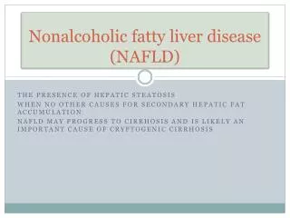
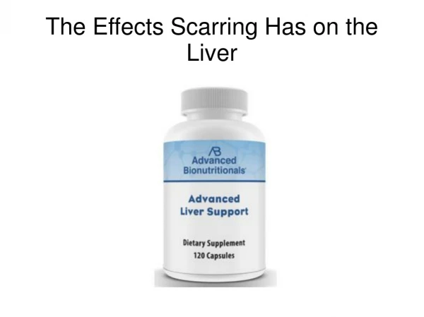
Non-alcohol fatty liver disease (NAFLD) Affects up to 85% of the obese • Defined as >5% of liver weight is from triacylglycerol • Associated with type II diabetes, hypertension, cardiovascular disease • Liver may make some of the TAG • Liver may be a dumping depot for TAG from other tissues • Precursor for steatohepatitis • Increased levels of AST and ALT (aminotransferase enzymes) NetNebraska.org stats: 55% of Nebraskans fit the “obese” category Least fit state in the Union 1 in 4 people in the world are overweight and now outnumber hungry people Qualityhealth.com: 10 fattest cities in America (Nov. 20, 2008) #7 – Scottsbluff, NE 31% obese, 9% T2D
Pathways to Liver illness Old Age Viral infection pseudocapillarization Capillarization of SEC & defenestration ECM in Space of Disse Activation of HSC Damage by high fat diet? Liver Fibrosis Increased ALT Cirrhosis What (cell) starts the cascade of events?
Overall Aims of project Measure liver SEC and HC stress as indicated by scavenger receptor function and fenestration in animals on specific fatty acid diets. 2. To determine the degree of ER stress in whole liver and nonparenchymal cells. 3. Determine cell-cell biochemical mediators that promote pro-inflammatory cellular activation Significance: This “disease” affects ~1/3 of the people in this state Innovation: No one has really looked at NAFLD from the SEC angle.
Aim 1 Measure liver SEC and HC stress as indicated by scavenger receptor function and fenestration in animals on specific fatty acid diets 60% Lard diet 12% Lard diet
Endocytosis Fenestration Purify SEC and HC from both rats • Hyaluronan for SEC • Orosomucoid for HC Young & healthy • 3 Methods that we can use • Injection of labeled ligand • Flush/excision of liver • Perfuse liver, isolate cells Old and sickly Visualize by SEM Porosity by liposomal transfer
Aim 2 ER stress Fatty acid overload Mitochondria Peroxisomes ROS ATF6 CHOP GRP78 XBP1 (cut) ER stress
Aim 2 Evaluation of ER stress in all 3 cell types: hepatocytes, stellate cells, SECs Perfuse livers of rats on both diets Purify cells Obtain mRNAs qRT-PCR: ATF6, CHOP, GRP78 PCR: XBP1 Use of cDNA arrays to fish for other markers
How liver cells talk to each other HSC HC Nitric oxide VEGF VEGF HGF SEC eNOS
How liver cells may talk to each other during NAFLD Activated HSC F-actin Timp1 a smooth muscle actin HC Nitric oxide ROS VEGF VEGF HGF Fibrosis SEC eNOS
Mode of action of the reactive oxygen species (ROS) ROS X eNOS IL-6 pathway Akt/PKB IKK NF-kB Increases in TIMP-1, aSMA, MMP-2, TGF-b and others?
Aim 3: Determine cell-cell biochemical mediators that promote pro-inflammatory cellular activation • 4 ways to quantify NO in liver • Whole tissue extract. cGMP is a marker for NO bioavailability. • eNOS converts L-Arg to NO and L-citrulline. Incubate purified SECs with • 3H-Arg and assays for 3H-citrulline. • Stain cells with 4-amino-5-methylamino-2’,7’-difluorofluorescein diacetate • (DAF-FM-DA; Molecular Probes) • 4. Co-cultures of activated HSC with SECs and look for response to quiescence.
ROS in hepatocytes in NAFLD vs control The principle reactive oxygen species we are interested in are superoxides (O2-) and peroxides H2O2 Hepatocytes from rats on each of the diets will be purified and plated. These can be measure by exposing cells to 2’7’-dichlorofluorescin diacetate
Cytokines and other pro-inflammatory mediators Whole Livers vs Purified Cells 2 primary methods: qRT-PCR (gene expression) and antibody-based kits (protein levels) Detect known culprits: TIMP-1, MMP-2, TGF-b Fish for others that may be cell specific: NF-kB array
Life at OU Proposed project SEC, HC purification SEC, HC culturing Endocytosis assays Rat injections Clearance strategies ELISA Enzyme assays Radiolabeling based assays RNA purification RT-PCR Fluorescence microscopy Liposome production MALLS Recombinant DNA Molecular biology Gene cloning Protein purification Stable cell line production Sugar modification SPR Mutagenesis Raising rats Cytokine measurements Nitric oxide chemistry Blood work Co-culturing HC, SEC HSC purification, culturing In vivo imaging qRT-PCR SEM