DNA Content Analysis of Synchronized BJ-EHLT Fibroblasts Using Hydroxyurea and RHPS4
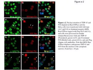
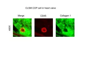
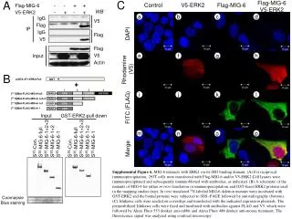
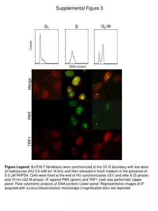
This study investigates the DNA content of BJ-EHLT fibroblasts synchronized at the G1-S boundary using a low dose of hydroxyurea (0.5 mM for 16 hours). Following the synchronization, cells were released into fresh medium containing 0.5 µM RHPS4. Cells were fixed at the end of the hydroxyurea treatment (G1 phase) and at 6 hours (S phase) and 10 hours (G2-M phase) post-release. Immunofluorescence against PAR (green) and TRF1 (red) was conducted. The upper panel displays flow cytometric analysis of DNA content, while the lower panel shows representative images obtained via a Leica Deconvolution microscope.

DNA Content Analysis of Synchronized BJ-EHLT Fibroblasts Using Hydroxyurea and RHPS4
E N D
Presentation Transcript
Supplemental Figure 3 G2-M G1 S Counts DNA content Merge PAR TRF1 Figure Legend: BJ-EHLT fibroblasts were synchronized at the G1-S boundary with low dose of hydroxiurea (HU 0.5 mM for 16 hrs) and then released in fresh medium in the presence of 0.5 M RHPS4. Cells were fixed at the end of HU synchronization (G1) and after 6 (S-phase) and 10 hrs (G2-M phase). IF against PAR (green) and TRF1 (red) was performed. Upper panel: Flow cytometric analysis of DNA content; Lower panel: Representative images of IF acquired with a Leica Deconvolution microscope (magnification 63x) are reported.