Merge
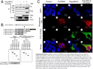
C. A. Flag-MIG-6 V5-ERK2. Control. V5-ERK2. Flag-MIG-6. Flag-MIG-6 - - + + V5-ERK2 - + - +. WB. a. b. c. d. IgG Flag IgG V5. V5 Flag Flag V5 Actin. IP. DAPI. Input. e. f. g. h. Rhodamine (V5). B. i. j. k. l.

Merge
E N D
Presentation Transcript
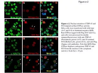
C A Flag-MIG-6 V5-ERK2 • Control • V5-ERK2 Flag-MIG-6 Flag-MIG-6 - - + + V5-ERK2 - + - + WB a b c d • IgG • Flag • IgG • V5 V5 Flag Flag V5 Actin IP DAPI Input e f g h • Rhodamine • (V5) B i j k l • FITC (FLAG) S35-Con. S35-MIG-6-full S35-MIG-6-1+2+3 S35-MIG-6-1+2 S35-MIG-6-1 S35-Con. S35-MIG-6-full S35-MIG-6-1+2+3 S35-MIG-6-1+2 S35-MIG-6-1 Input GST-ERK2-pull down m n o p • Merge Supplemental Figure 6.MIG-6 interacts with ERK2 via its SH3 binding domain. (A) For reciprocal immunoprecipitation, 293T cells were transfected with Flag-MIG-6 and/or V5-ERK2.Cell lysates were immunoprecipitated and subsequently immunoblotted with antibodies, as indicated. (B) A schematic of the mutants of MIG-6 for either in vitro translation or immunoprecipitation and GST-fused ERK2 proteins used in the mapping studies (top). In vitro translated 35S-labeled MIG-6 deletion mutants were incubated with GST-ERK2 and the bound proteins were subjected to SDS–PAGE followed by autoradiography (bottom). (C) Ishikawa cells were seeded on coverslips and transfected with the indicated expression plasmids. The permeabilizedIshikawa cells were fixed and incubated with antibodies against FLAG and V5, which were followed by Alexa Fluor 555 donkey anti-rabbit and Alexa Fluor 488 donkey anti-mouse treatment. The fluorescence signal was analyzed using confocal microscopy. Coomassie Blue staining