Download
1 / 49
490 likes | 514 Vues
Explore the key components and steps of the human immune system's defense mechanisms against pathogens and infections. Learn about barriers, cell responses, inflammation, and antibody-mediated and cell-mediated immunity.

E N D
Three Lines of Defense • Barriers at body surfaces • Nonspecific responses • Immune responses
Cells Involved in Immune Responses basophil mast cell eosinophil neutrophil NK cell B lymphocyte T lymphocyte Fig. 39-3a, p.681
Cells Involved in Immune Responses dendritic cell macrophage Fig. 39-3b, p.681
Barriers at Body Surface • Intact skin and mucous membranes • Lysozyme • Normal bacterial flora • Flushing effect and low pH of urine
Barriers Against Infectious Agents Fig. 39-6, p.683
Barriers Against Infectious Agents Fig. 39-7, p.683
Nonspecific Responses • Lymph nodes trap and kill pathogens • Natural killer cells attack a range of targets • Inflammation
Complement System • Plasma proteins that take part in both specific and nonspecific response • Activation of one triggers cascade of reactions that activate others
Attack Complexes lipid bilayer of pathogen antibody activated complement bacterial pathogen Cascade Reactions Formation of Attack Complexes Lysis of Target Activation
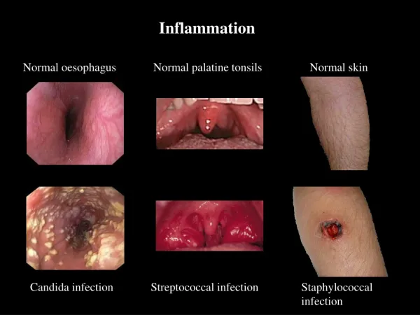
Acute Inflammation • Nonspecific response to foreign invasion, tissue damage, or both • Destroys invaders, removes debris, and prepares area for healing • Characterized by redness, swelling, warmth, and pain
Inflammation • Mast cells release histamine • Capillaries dilate and leak • Complement proteins attack bacteria • White cells attack invaders and clean up Figure 39.9Page 685
Features of Immune Responses • Self/nonself recognition • Specificity • Diversity • Memory
Memory and Effector Cells • When a B or T cell is stimulated to divide, it produces more than one cell type • Memory cells - set aside for future use • Effector cells - engage and destroy the current threat
Steps in Immune Response • Recognition of an antigen • Rounds of cell division that form huge populations of lymphocytes • Specialization of lymphocytes into effector and memory cells that have receptors for one kind of antigen
Key Components of Immune Response • MHC markers • Antigen-presenting cells • T cells • B cells • Natural killer cells
Formation of Antigen-MHC Complex antigen fragments MHC molecule antigen-MHC complex
Antigens • “Nonself” markers on foreign agents and altered body cells such as tumors • Trigger division of B and T cells
Lymphocyte Battlegrounds • Lymph nodes filter antigens from body fluids • Macrophages, dendritic cells, B and T cells in nodes and spleen mount a defense Figure 39.12Page 687
Antibody Structure • Consists of four polypeptide chains • Certain parts of each chain are variable; impart antigen specificity antigen binding site variable region constant region Figure 39.13aPage 688
signal reception signal transduction cellular response Antibody-Mediated Immune Response Cell-Mediated Immune Response antigen-presenting cells naive B cells + antigen + complement naive helper T cells activated B cells naive cytotoxic T cells effector helper T cells + memory helper T cells effector B cells + memory B cells effector cytotoxic T cells + memory cytotoxic cells Fig. 39-11, p.687
Antibody-Mediated Response • Carried out by B cells • Targets are intracellular pathogens and toxins • Antibodies bind to target and mark it for destruction by phagocytes and complement
antigen Clonal Selection • Only the B cell with antigen-receptor that matches antigen is stimulated to divide • Mitosis yields many cells with that receptor clonal population of B cells Figure 39.15Page 690
Immunological Memory • Memory cells specific for an antigen are quickly activated to divide upon subsequent exposure to that antigen Primary Immune Response: naïve T or B cell effector cells memory cells Secondary Immune Response: effector cells memory cells Figure 39.15Page 690
Primary and Secondary Immune Response Fig. 39-16, p.690
Antibody-Mediated Response • B cell becomes antigen-presenting cell • Helper T cell binds to antigen-MHC complex • Interleukins stimulate B cell division and differentiation • Effector cells secrete antibodies antigen naïve B cell MHC molecule antigen- MHC complex helper T cell antigen- presenting B cell interleukins memory B cell antibody effector B cell
bacterium dendritic cell complement naive B cell naive T cell antigen-presenting cell cytokines memory helper T cell effector helper T cell B cell effector B cell memory B cell Stepped Art Fig. 39-17, p.691
virus particle (red) infecting a body cell (yellow) dendritic cell antigen-MHC complex naive cytotoxic T cell naive helper T cell antigen-presenting cell effector cytotoxic T cell memory cytotoxic T cell effector cytotoxic T cell cytokines memory helper T cell effector helper T cell activated cytotoxic T cell Stepped Art Fig. 39-18, p.692
Cell-Mediated Response another macrophage one macrophage • Carried out by T cells • Stimulated by antigen-presenting macrophages • Main target is antigen-presenting body cells (cells with intracellular pathogens) or tumor cells interleukins cytotoxic T cell helper T cell interleukins infected body cell
Cytotoxic T Cell cytotoxic T-cell tumor cell Fig. 39-19, p.693
Organ Rejection • Cytotoxic T cells can contribute to rejection • They recognize a portion of the donor cell’s MHC complex as self, view a portion as foreign • Treat the combination as an antigen-MHC complex and attack donor cells
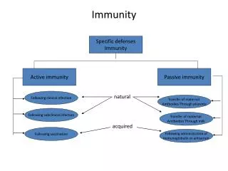
Immunization • Process that promotes immunity • Active immunization - • Antigen-containing material is injected • Confers long-lasting immunity • Passive - • Purified antibody is injected • Protection is short lived
Vaccines Fig. 39-21a, p.694
Vaccines RECOMMENDED VACCINES RECOMMENDED AGES Hepatitis B Hepatitis B booster 1 Hepatitis B booster 2 Hepatitis B assessment DTP (Diphtheria; Tetanus; and Pertussin, or whooping cough) DTP booster 1 DTP booster 2 DT HiB (Hemophilus influenzae) HiB booster Polio Polio booster 1 Polio booster 2 MMR (Measles, Mumps, Rubella) MMR booster MMR assessment Pneumococcal Pneumococcal booster 1 Pneumococcal booster 2 Varicella Varicella assessment Hepatitis A (in selected areas Birth–2 months 1–4 months 6–18 months 11–12 years 2, 4, and 6 months 15–18 months 4–6 years 11–16 years 2, 4, and 6 months 12–15 months 2 and 4 months 6–18 months 4–6 years 12–15 months 4–6 years 11–12 years 2, 4, and 6 months 12-15 months 1-18 years 12–18 months 11–12 years 1-12 years Fig. 39-21b, p.694
Allergies • Immune reaction to a harmless substance • Genetic predisposition • IgE responds to antigen by binding to mast cells and basophils • These cells secrete the substances that cause symptoms
AIDS • Combination of disorders that follows infection with HIV • Includes • Yeast (Candida) infections • Pneumocystis pneumonia • Karposi’s sarcoma
HIV Life Cycle reverse transcriptase viral genes are integrated into the host DNA DNA is transcribed viral RNA enters cell host cell strands of DNA (two) viral RNA reverse transcription of viral RNA viral proteins viral DNA budding core proteins (two layers) integrase
T Cell Decline • Release of new viral particles kills the host T cell • The body is constantly making new T cells, but cannot outpace the rate of destruction • As infection proceeds, T cell numbers inevitably decline
Effect of T Cell Decline • CD4 helper T cells play a vital role in immune function • They are required for both cell-mediated and antibody-mediated immunity • Infected individual becomes vulnerable to other infections, which eventually result in death