The Anaerobes
The Anaerobes. Bacteriodaceae, Clostridium, and the anaerobic cocci. Bacteriodaceae. Classification Bacteroidaceae family includes the following genera Bacteroides Fusobacterium Leptotrichia (rare in human diseases) Prevotella Porphyromomas

The Anaerobes
E N D
Presentation Transcript
The Anaerobes Bacteriodaceae, Clostridium, and the anaerobic cocci
Bacteriodaceae • Classification • Bacteroidaceae family includes the following genera • Bacteroides • Fusobacterium • Leptotrichia (rare in human diseases) • Prevotella • Porphyromomas • B. fragilis is the most commonly isolated anaerobic G-B. • All are nonsporing, anaerobic, G-B
Bacteriodaceae • They may be NF of the oropharynx, urogenital tract, and colon and are considered opportunistic pathogens • Morphology/cultural characteristics • Pleomorphic G-B showing irregular or bipolar staining. • Fusobacterium nucleatum characteristically is long and slender with pointed ends
Bacteriodaceae • To grow these organisms, nonselective anaerobic BA plates, selective anaerobic plates, and liquid should be used for primary isolation. • Nonselective anaerobic BA plates= CBA plates plus vitamin K1, hemin, yeast extract, and L-cystine (supplemented BA) • Selective media (all for Bacteroides species) • Anaerobic PEA BA – suppresses aerobic G-B • Kanamycin-Vancomycin BA – inhibits G+ and facultatively anaerobic G -
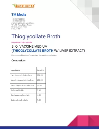
Bacteriodaceae • Kanamycin-Vancomycin laked BA (KVLB) prepared by freezing and thawing whole blood • Bacteroides Bile Esculin (BBE) agar • Liquid media • Thioglycollate • Chopped meat glucose • Must incubate under strict anaerobic conditions • Incubation at 35-370 C for 48 hours before opening an anaerobic jar.
Bacteriodaceae • Each colony type that grows should be Gram stained and subcultured to plates grown under both aerobic and anaerobic conditions to confirm that it is an anaerobe. • Biochemistry • The Bacteroides group which now includes Prevotella and Porphyromonas species are divided into groups based on bile tolerance, pigment production, and sensitivity to the antibiotics Vancomycin (V), Kanamycin (K), and Colistin (C)
Bacteriodaceae V K C Bile pigment Bacteroides fragilis R R R R - Prevotella R R S S +/- Porphyromonas S R R S + • B. fragilis is catalase + • GLC – used to differentiate Fusobacterium from the others. • The major by-product of Fusobacterium is butyric acid while the others produce mixtures of acids. • F. nucleatum and F. necrophorum (lipase+) are the major pathogens
Bacteriodaceae • Virulence factors • Fusobacterium – endotoxin; the endotoxin of Bacteroides is not highly toxic • B. fragilis – capsule • Some in the Bacteroides group produce IgA protease,collagenase, phosphotase, RNAse, or DNAse • Clinical significance
Bacteriodaceae • Clinical significance • These organisms are NF of the oropharynx, urogenital tract, and colon and cause serious infections when they gain access to tissues and organs. • Most commonly they cause intra-abdominal infections • The infections are usually polymicrobial infections • They can also be found causing: • Peridontal disease
Bacteriodaceae • Chronic otitis media • Chronic sinusitis • Wound infections • Pneumonia • Female genital tract infections • Brain abscesses • Bacterial endocarditis • Bone infections • Antimicrobial susceptability/treatment • Incision and drainage • Chloramphenicol, clindamycin, cefoxitin, or metronidazole
Clostridium • Classification – no family designation • Most are strict anaerobes • Are widely distributed in soil and water • Some are NF in the GI tract of man and other animals • Morphology/cultural characteristics • Are endospore forming large G+B • The site at which the endospore forms in the vegetative cell is characteristic and helps in differentiating C. tetani (terminal) from the others (oval and subterminal)
Clostridium • All except C. perfringens are motile • Nonselective, selective, and liquid media should be used for primary isolation • Nonselective – supplemented anaerobic BA • C. perfringens produces a classic double zone of hemolysis • Nonselective, differential – Egg yolk agar • Allows differentiation based on • Lecithinase production (white precipitate) • Lipase production (sheen around surface of colonies) • Protease production(clearing)
Clostridium • Selective – Cycloserine-cefoxitin-egg yolk-fructose agar(CCFA) is selective for C. difficile • Liquid • Thioglycollate • Chopped meat • Special isolation procedures – Clostridia usually occur in mixed cultures with G-B and nonsporing anaerobes – use heat or alcohol treatment to kill others before plating • C. perfringens grows rapidly at 450 C
Clostridium • Biochemistry • O2 tolerance – C. tertium and C. histolyticum are aerotolerant, but catalase - • Lipase vs lecithinase vs protease production on egg yolk agar • Naegler reaction - smear ½ of an egg yolk agar plate with type A anti-toxin (anti-lecithinase), streak organism in a single line, and look for inhibition of lecithinase production • Sugar fermentations
Clostridium • Milk digestion • Esculin hydrolysis • Gelatin hydrolysis • Reverse CAMP – is presumptive for C. perfringens • Mechanisms of Virulence – most Clostridia are not invasive, but many produce powerful toxins and enzymes • C. perfringens produces a capsule • C. botulinum – produces a potent exotoxin • The organisms are divided into 8 different types based on which of the 8 serotypes of exotoxin are produced.
Clostridium • Serotypes are A, B, C1, C2, D, E, F, and G • Serotype A is the most potent. • Types A,B, E, F, and G can cause botulism in man • Botulinal exotoxin is the most powerful exotoxin known. • It works at the neuromuscular junction and in the autonomic nervous system to prevent the release of the neurotransmitter acetylcholine. • This leads to flaccid paralysis. • The toxin has two components, B for binding, and A for the toxic activity. • The toxic part cleaves proteins that mediate fusion of synaptic vesicles with the cell membrane and subsequent release of acetylcholine. • The toxin is part of the bacteria and not released until the death of the bacteria.
Proteins involved in membrane fusion to release acetylcholine
Clostridium • C. tetani -produces two exotoxins • A hemolysin • Tetanospasmin – can travel to the CNS humorally through blood and lymph, or neurally through tissue spaces of the peripheral nerves. • The toxin binds to sialic acid containing gangliosides of the CNS to prevent the release of the inhibitory neurotransmitters GABA and glycine from synapses (by cleaving VAMP) in the inhibitory nerve system of the spinal cord. • This is a system that prevents the contraction of a muscle when the muscle of the opposite action contracts. • This leads to both sets of muscles contracting at the same time and spastic paralysis. • Called lockjaw when the jaw is affected. • Contractions can break the back.
Clostridium • C. perfringens – is divided into 5 types, A through E, based on the major lethal toxins produced. • All types produce the alpha toxin – is a lethal, necrotizing lecithinase which is responsible for the outer zone of hemolysis. • Other toxins that may be produced include beta, epsilon, or iota which are all lethal and necrotizing, delta and theta which are lethal and hemolytic and are responsible for the inner zone of hemolysis.
Clostridium • Enzymes produces may include gelatinase, collagenase, protease, hyaluronidase, DNAse, and neuraminidase • Types A and C produce an enterotoxin responsible for causing an intoxication type of food poisoning in meats, poultry, and gravy. • Its action resembles that of the cholera toxin.
Clostridium • C. difficile – produces two exotoxins both of which inactivate Rho proteins by adding a glucose. • Rho proteins function as molecular switches in cytoskeletal dynamics and many signal transduction pathways • Enterotoxin A stimulates fluid and electrolyte losses from the intestinal tract • Cytotoxin B kills mammalian cells
Clostridium • Clinical significance • C. tetani – causes tetanus, a disease due to the toxin tetanospasmin. • Spores are found in feces, soil, and dust. Spores enter the body through wounds where they germinate into vegetative cells and subsequently produce toxin when a sufficiently low O/R potential is established in the infected tissue (usually a deep wound).
Clostridium • The incubation is 1-54 days with an average of 6-15 days. The longer the incubation, the better the prognosis. • Symptoms begin with cramps and twitching of muscles around the wound. • Headache and neck stiffness also occur. • These are followed by trismus (lockjaw) and more generalized symptoms. • Death, if it occurs, results from respiratory failure within 4 days. • Neonatal tetanus is a consequence of infection of the umbilicus through septic midwifery and it occurs in underdeveloped countries.
Clostridium • C. botulinum – causes botulism • Food botulism – in the U.S. this usually occurs following ingestion of inadequately processed home-canned food. For this to occur you need: • Food must be contaminated with C. botulinum spores • Food must possess composition and nutritive properties that allow germination and toxin production • Food must have suitable pH and temperature • Food must have been inadequately heated or processed (toxin is heat labile)
Clostridium • Following ingestion, toxin is absorbed from the intestine and transported via blood and lymph to the PNS. • The incubation is 8 hours to 8 days with 18-36 hours most common • The first symptoms include nausea, vomiting, and diarrhea followed by symmetric, descending paralysis (eyes, throat, neck, trunk, and then the limbs) • Paralysis of respiratory muscles results in death • Infant botulism – follows ingestion of spores which germinate in the intestine.
Clostridium • Illness may range from subclinical to sudden infant death syndrome • Honey has been implicated as a source of spores. • This doesn’t occur in adults because of competing NF • Wound botulism – can follow C. botulinum toxin production in a traumatic wound. • Clostridial wound infections – most species of Clostridium are saprophytic bacteria living in soil and water. • C. perfringens and other are found in the intestines of man and other animals.
Clostridium • Most clostridial wound infections occur as simple contaminants of a fresh wound and most heal normally with simple therapy • Anaerobic cellulitis follows invasion of necrotic wound tissue by proteolytic bacteria and is characterized by gas accumulation, discoloration of the skin, and a malodorous brown, purulent discharge. • Clostridial myonecrosis or gas gangrene – involves invasion of normal, healthy muscle tissue surrounding the wound site. • C. perfringens is most commonly isolated. Need a lowered O/R potential in the wound which causes reduction of pyruvate to lactate and a decreased pH which activates the proteolytic enzymes. • Clinical features include severe systemic toxicity, a painful, edematous wound with a sweet or foul smelling discharge. • Untreated cases may result in death.
Clostridium • C. difficile – Antibiotic therapy of any kind can result in diarrhea. • The severity may range from simple diarrhea to severe antibiotic associated pseudomembranous colitis. • This is characterized by colonic plaques that coalesce to form a pseudomembrane of mucin, fibrin, sloughed off epithelial cells, and acute inflammatory cells. • Complications include dehydration, electrolyte imbalance and colonic perforation. • Most often occurs following therapy with ampicillin, clindamycin, or cephalosporins and is usually due to C. difficile which may be NF in the G.I. tract.