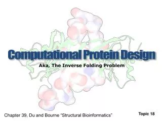
Computational design of protein function
200 likes | 369 Vues
Computational design of protein function. Loren Looger Hellinga lab. 1. Allowable structures for proteins, DNA, small molecules. Progesterone. 2. Pseudo-geometric potential. electrostatics. H-bonds. sterics. solvation. Pretty much like CHARMM. a ~ 1.1. E. r. Hydrogen bonds, too.

Computational design of protein function
E N D
Presentation Transcript
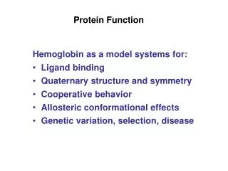
Computational design of protein function Loren Looger Hellinga lab
1. Allowable structures for proteins, DNA, small molecules Progesterone
2. Pseudo-geometric potential electrostatics H-bonds sterics solvation
Pretty much like CHARMM... a ~ 1.1 E r
Hydrogen bonds, too... anchor r H A D { } -8 · · ·
Area-based solvation energy P P P = polar H = hydrophobic H H H
Electrostatic potential is a function of atom-type pair & protein environment. Parameterized to fit experimental data.
3. Algorithm for choosing best structure(s) from all available
Complementary Surface Construction: Protein coordinates Ligand coordinates Poly-alanine PCS Rotational ligand ensemble Docking grid Force field Placed ligand ensemble Fixed ligand ensemble Side-chain rotamers Evolved PCS ensemble Ranked PCS ensemble Experiments Periplasmic Binding Protein (PBP) scaffolds Molten zone Evolving zone Fixed zone
Metabolites 1 F D-lactate & L-lactate Kd = 2 µM x 0.5 1 1 1 0 40 80 serotonin [L-lactate] (µM) F Kd = 6 µM x F 5-fluorouracil Kd = 4 nM x 0.5 0.5 0.5 0 0 52.5 100 0 [serotonin] (µM) 0 150 300 [5-fluorouracil] (nM) ibuprofen dopamine 1 F F x x Kd = 6 µM Kd = 2 nM 0.5 MTBE 1 F 0 x 50 100 12.5 25 RDX TNT [MTBE] (µM) Kd = 45 nM [TNT] (nM) 0.5 PMPA~soman 0.25 0.5 [PMPA] (µM) 0 Neurotransmitters Drugs Pollutants Explosives Chemical Threats
QSAR Results for binding affinities for L-lactate & TNT Receptors -8 -6 log Kd (obs) -4 -2
QBP 100mM 10 RBP 1 100µM 10 1 0.1 HBP ABP L-lactate designs GBP
QBP 100mM 10 RBP 1 100µM 10 1 0.1 HBP ABP The use of QSARs in the predictions improves the designs: D-lactate GBP
Construction of biological sentinels for chemical threats and pollutants binary modulation expression [inducer]
Unicellular sentinels for chemical threats and pollutants - + - + TNT Ribose Lactate MTBE 5 Fluoro-uracil
Dose Response of TNTa Signaling IPTG 0 mM TNT 100 mM 2,4-DNT 100 mM 2,6-DNT 100 mM 10 mM 1 mM 0.1 mM 0.01 mM 0.001 mM [TNT]
Kdlactate Racemic mix D L none none 200µM 3µM 0.8µM 10µM Absorbance 210nm D L Optically pure enantiomers D L Wt Gbp Immobilized receptors L-Lac.G1 D-Lac.G1 Fraction #
Calculation #2 Complementary surface construction (PCS + SCS) geometrical description of essential features in the complementary surface Calculation #1 Initial placement of PCS on scaffold backbone { l, w1, w2, q1, q2, q3 }n Combinatorial search (108 sequence 1012 rotamers) Complementary surface construction (1010-10200 rotamers) Site 1 Site 1 Design scaffold coordinates side-chain rotamer library Site 2 Site 2 +... +... Pairwise of atomic interactions Computational design of ligand-binding sitesStrategy #2: predefined geometries
Acknowledgements • Mary Dwyer • Jeff Smith • Shahir Rizk