Protein function
This lecture delves into the intricacies of protein function, focusing on domain fusion within bioinformatics. It discusses the multi-enzyme protein present in vertebrates, comprising GARs, AIRs, and GARt enzymes. It highlights genetic mechanisms influencing multidomain protein structure and the concept of domain swapping as a method for forming oligomeric assemblies. Additionally, it introduces the Clusters of Orthologous Groups (COGs) database and the PRINTS database for identifying conserved motifs in protein families, providing a framework for functional and evolutionary genome analysis.

Protein function
E N D
Presentation Transcript
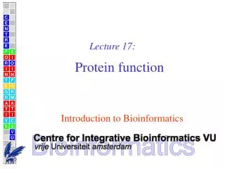
C E N T E R F O R I N T E G R A T I V E B I O I N F O R M A T I C S V U Lecture 17: Protein function Introduction to Bioinformatics
Domain fusion For example, vertebrates have a multi-enzyme protein (GARs-AIRs-GARt) comprising the enzymes GAR synthetase (GARs), AIR synthetase (AIRs), and GAR transformylase (GARt) 1. In insects, the polypeptide appears as GARs-(AIRs)2-GARt. However, GARs-AIRs is encoded separately from GARt in yeast, and in bacteria each domain is encoded separately (Henikoff et al., 1997). 1GAR: glycinamide ribonucleotide synthetase AIR: aminoimidazole ribonucleotide synthetase
Domain fusion Genetic mechanisms influencing the layout of multidomain proteins include gross rearrangements such as inversions, translocations, deletions and duplications, homologous recombination, and slippage of DNA polymerase during replication (Bork et al., 1992). Although genetically conceivable, the transition from two single domain proteins to a multidomain protein requires that both domains fold correctly and that they accomplish to bury a fraction of the previously solvent-exposed surface area in a newly generated inter-domain surface.
Domain swapping Domain swapping is a structurally viable mechanism for forming oligomeric assemblies (Bennett et al., 1995). In domain swapping, a secondary or tertiary element of a monomeric protein is replaced by the same element of another protein. Domain swapping can range from secondary structure elements to whole structural domains. It also represents a model of evolution for functional adaptation by oligomerization, e.g. of oligomeric enzymes that have their active site at sub-unit interfaces (Heringa and Taylor, 1997).
COGS Domain database The COGs (Clusters of Orthologous Groups) database is a phylogenetic classification of the proteins encoded within complete genomes (Tatusov et al., 2001). It primarily consists of bacterial and archaeal genomes. Operational definition of orthology is based on bidirectional best hit Incorporation of the larger genomes of multicellular eukaryotes into the COG system is achieved by identifying eukaryotic proteins that fit into already existing COGs. Eukaryotic proteins that have orthologs within different COGs are split into their individual domains. The COGs database currently consists of 3166 COGs including 75,725 proteins from 44 genomes.
COGs: the beginning (1997) In order to extract the maximum amount of information from the rapidly accumulating genome sequences, all conserved genes need to be classified according to their homologous relationships. Comparison of proteins encoded in seven complete genomes from five major phylogenetic lineages and elucidation of consistent patterns of sequence similarities allowed the delineation of 720 clusters of orthologous groups (COGs). Each COG consists of individual orthologous proteins or orthologous sets of paralogs from at least three lineages. Orthologs typically have the same function, allowing transfer of functional information from one member to an entire COG. This relation automatically yields a number of functional predictions for poorly characterized genomes. The COGs comprise a framework for functional and evolutionary genome analysis.
COG2813:16S RNA G1207 methylase RsmC COG members are mapped onto the genomes included in the DB
PRINTS database • PRINTS is a compendium of protein fingerprints. • A fingerprint is a group of conserved motifs used to characterise a protein family; its diagnostic power (false positives and false negatives) is refined by iterative scanning of a SWISS-PROT/TrEMBL composite database. • Usually the motifs do not overlap, but are separated along a sequence, though they may be contiguous in 3D-space. • Fingerprints can encode protein folds and functionalities more flexibly and powerfully than can single motifs, full diagnostic potency deriving from the mutual context provided by motif neighbours • PRINTS contains the most discriminating groups of regular expressions for each protein sequence • Release 31.0 of PRINTS contains 1,550 entries, encoding 9,531 individual motifs.
BETAHEAM: 2 of 5 PRINTS motifs making the fingerprint INITIAL MOTIF SETS BETAHAEM1 Length of motif = 17 Motif number = 1 Beta haemoglobin motif I - 1 PCODE ST INT GRLLVVYPWTQRYFDSF HBB1_RAT 29 29 GRLLVVYPWTQRYFDSF HBB1_MOUSE 29 29 GRLLVVYPWTQRFFEHF HBB_ALCAA 28 28 GRLLVVYPWTQRFFEHF HBB_ODOVI 28 28 GRLLVVYPWTQRFFESF HBB_BOVIN 28 28 GRLLVVYPWTQRFFESF HBB_ATEGE 29 29 GRLLVVYPWTQRFFESF HBB_HUMAN 29 29 GRLLVVYPWTQRFFESF HBB_ANTPA 29 29 ARLLIVYPWTQRFFASF HBB_ANAPL 29 29 SRCLIVYPWTQRHFSGF HBB_NOTAN 29 29 BETAHAEM2 Length of motif = 16 Motif number = 2 Beta haemoglobin motif II - 1 PCODE ST INT DLSSASAIMGNPKVKA HBB1_RAT 47 1 DLSSASAIMGNAKVKA HBB1_MOUSE 47 1 DLSTADAVMHNAKVKE HBB_ALCAA 46 1 DLSSAGAVMGNPKVKA HBB_ODOVI 46 1 DLSTADAVMNNPKVKA HBB_BOVIN 46 1 DLSTPDAVMSNPKVKA HBB_ATEGE 47 1 DLSTPDAVMGNPKVKA HBB_HUMAN 47 1 DLSNAGAVMGNAKVKA HBB_ANTPA 47 1 NLSSPTAILGNPMVRA HBB_ANAPL 47 1 NLYNAEAILGNANVAA HBB_NOTAN 47 1 After iteration the number of sequences for each motif can grow dramatically. Both the initial motifs (example here) and final motifs are provided to the user
The PRODOM Database ProDom is a comprehensive set of protein domain families automatically generated from the SWISS-PROT and TrEMBL sequence databases
The PRODOM Database ProDom (Corpet et al., 2000) is a database of protein domain families automatically generated from SWISSPROT and TrEMBL sequence databases (Bairoch and Apweiler, 2000) using a novel procedure based on recursive PSI-BLAST searches (Altschul et al., 1997). Release 2001.2 of ProDom contains 283,772 domain families, 101,957 having at least 2 sequence members. ProDom-CG (Complete Genome) is a version of the ProDom database which holds genome-specific domain data.
The PROSITE Database PROSITE is a database of protein families and domains. It consists of biologically significant sites, patterns and profiles that help to reliably identify to which known protein family (if any) a new sequence belongs PROSITE (Hofmann et al., 1999) is a good source of high quality annotation for protein domain families. A PROSITE sequence family is represented as a pattern or profile, providing a means of sensitive detection of common protein domains in new protein sequences. PROSITE release 16.46 contains signatures specific for 1,098 protein families or domains. Each of these signatures comes with documentation providing background information on the structure and function of these proteins.
The PROSITE Database A PROSITE sequence family is represented as a pattern or a profile. A pattern is given as a regular expression (next slide) The generalised profiles used in PROSITE carry the same increased information as compared to classical profiles as Hidden Markov Models (HMMs).
Regular expressions Alignment ADLGAVFALCDRYFQ SDVGPRSCFCERFYQ ADLGRTQNRCDRYYQ ADIGQPHSLCERYFQ Regular expression [AS]-D-[IVL]-G-x4-{PG}-C-[DE]-R-[FY]2-Q {PG} = not (P or G) For short sequence stretches, regular expressions are often more suitable to describe the information than alignments (or profiles)
Regular expressions Regular expression No. of exact matches in DB D-A-V-I-D 71 D-A-V-I-[DENQ] 252 [DENQ]-A-V-I-[DENQ] 925 [DENQ]-A-[VLI]-I-[DENQ] 2739 [DENQ]-[AG]-[VLI]2-[DENQ] 51506 D-A-V-E 1088
Rationale for regular expressions • “I want to see all sequences that ... • ... contain a C” --- C • ... contain a C or an F” -- [CF] • ... contain a C and an F” -- (C.*F | F.*C) (‘|’ means ‘or’ and ‘.*’ means don’t care for any length) • ... contain a C immediately followed by an F” -- CF • ... contain a C later followed by an F” -- C.*F • ... begin with a C” -- ^C (‘^’ means ‘starting with’) • ... do not contain a C” -- {C} • ... contain at least three Cs” -- C3- • ... contain exactly three Cs” -- C3 • ... has a C at the seventh position” -- .6C • ... either contain a C, an E, and an F in any order except CFE, unless there are also at most three Ps, or there is a ....
Regex limitations • regex cannot remember indeterminate counts !!! • “I want to see all sequences with ... • ... six Cs followed by six Ts” • C6T6 • ... any number of Cs followed by any number of Ts” • C*T* • ... Cs followed by an equal number of Ts” (This cannot be done..) • CnTn • (CT|CCTT|CCCTTT|C4T4| ... )?
The PFAM Database Pfam is a large collection of multiple sequence alignments and hidden Markov models covering many common protein domains and families. For each family in Pfam you can: • Look at multiple alignments • View protein domain architectures • Examine species distribution • Follow links to other databases • View known protein structures • Search with Hidden Markov Model (HMM) for each alignment
The PFAM Database Pfam is a database of two parts, the first is the curated part of Pfam containing over 5193 protein families (Pfam-A). Pfam-A comprises manually crafted multiple alignments and profile-HMMs . To give Pfam a more comprehensive coverage of known proteins we automatically generate a supplement called Pfam-B. This contains a large number of small families taken from the PRODOM database that do not overlap with Pfam-A. Although of lower quality Pfam-B families can be useful when no Pfam-A families are found.
The PFAM Database Sequence coverage Pfam-A : 73% (Gr) Sequence coverage Pfam-B : 20% (Bl) Other (Grey)
A PFAM alignment CYB_TRYBB/1-197 M...LYKSG..EKRKG..LLMSGC.....LYR.....IYGVGFSLGFFIALQIIC..GVCLAWLFFSCFICSNWYFVLFLCYB_MARPO/1-208 M.ARRLSILKQPIFSTFNNHLIDY.....PTPSNISYWWGFGSLAGLCLVIQILTGVFLAMHYTPHVDLAFLSVEHIMR.CYB_HETFR/1-205 MATNIRKTH..PLLKIINHALVDL.....PAPSNISAWWNFGSLLVLCLAVQILTGLFLAMHYTADISLAFSSVIHICR.CYB_STELO/1-204 M.TNIRKTH..PLMKILNDAFIDL.....PTPSNISSWWNFGSLLGLCLIMQILTGLFLAMHYTPDTTTAFSSVAHICR.CYB_ASCSU/1-196 ...........MKLDFVNSMVVSL.....PSSKVLTYGWNFGSMLGMVLGFQILTGTFLAFYYSNDGALAFLSVQYIMY.CYB6_SPIOL/1-210 M.SKVYDWF..EERLEIQAIADDITSKYVPPHVNIFYCLGGITLT..CFLVQVATGFAMTFYYRPTVTDAFASVQYIMT.CYB6_MARPO/1-210 M.GKVYDWF..EERLEIQAIADDITSKYVPPHVNIFYCLGGITLT..CFLVQVATGFAMTFYYRPTVTEAFSSVQYIMT.CYB6_EUGGR/1-210 M.SRVYDWF..EERLEIQAIADDVSSKYVPPHVNIFYCLGGITFT..CFIIQVATGFAMTFYYRPTVTEAFLSVKYIMN.CYB_TRYBB/1-197 WDFDLGFVIRSVHICFTSLLYLLLYIHIFKSITLIILFDTH..IL....VWFIGFILFVFIIIIAFIGYVLPCTMMSYWGCYB_MARPO/1-208 .DVKGGWLLRYMHANGASMFFIVVYLHFFRGLY....YGSY..ASPRELVWCLGVVILLLMIVTAFIGYVLPWGQMSFWGCYB_HETFR/1-205 .DVNYGWLIRNIHANGASLFFICIYLHIARGLY....YGSY..LLKE..TWNIGVILLFLLMATAFVGYVLPWGQMSFWGCYB_STELO/1-204 .DVNYGWFIRYLHANGASMFFICLYAHMGRGLY....YGSY..MFQE..TWNIGVLLLLTVMATAFVGYVLPWGQMSFWGCYB_ASCSU/1-196 .EVNFGWIFRVLHFNGASLFFIFLYLHLFKGLF....FMSY..RLKK..VWVSGIVILLLVMMEAFMGYVLVWAQMSFWACYB6_SPIOL/1-210 .EVNFGWLIRSVHRWSASMMVLMMILHVFRVYL....TGGFKKPREL..TWVTGVVLGVLTASFGVTGYSLPWDQIGYWACYB6_MARPO/1-210 .EVNFGWLIRSVHRWSASMMVLMMILHIFRVYL....TGGFKKPREL..TWVTGVILAVLTVSFGVTGYSLPWDQIGYWACYB6_EUGGR/1-210 .EVNFGWLIRSIHRWSASMMVLMMILHVCRVYL....TGGFKKPREL..TWVTGIILAILTVSFGVTGYSLPWDQVGYWACYB_TRYBB/1-197 LTVFSNIIATVPILGIWLCYWIWGSEFINDFTLLKLHVLHV.LLPFILLIILILHLFCLHYFMCYB_MARPO/1-208 ATVITSLASAIPVVGDTIVTWLWGGFSVDNATLNRFFSLHY.LLPFIIAGASILHLAALHQYGCYB_HETFR/1-205 ATVITNLLSAFPYIGDTLVQWIWGGFSIDNATLTRFFAFHF.LLPFLIIALTMLHFLFLHETGCYB_STELO/1-204 ATVITNLLSAIPYIGTTLVEWIWGGFSVDKATLTRFFAFHF.ILPFIITALAAVHLLFLHETGCYB_ASCSU/1-196 SVVITSLLSVIPVWGFAIVTWIWSGFTVSSATLKFFFVLHF.LVPWGLLLLVLLHLVFLHETGCYB6_SPIOL/1-210 VKIVTGVPDAIPVIGSPLVELLRGSASVGQSTLTRFYSLHTFVLPLLTAVFMLMHFLMIRKQGCYB6_MARPO/1-210 VKIVTGVPEAIPIIGSPLVELLRGSVSVGQSTLTRFYSLHTFVLPLLTAIFMLMHFLMIRKQGCYB6_EUGGR/1-210 VKIVTGVPEAIPLIGNFIVELLRGSVSVGQSTLTRFYSLHTFVLPLLTATFMLGHFLMIRKQG
INTERPRO combined database Because the underlying construction and analysis methods of the above domain family databases are different, the databases inevitably have different diagnostic strengths and weaknesses. The InterPro database (Apweiler et al., 2000) is a collaboration between many of the domain database curators. It aims to be a central resource reducing the amount of duplication between the databases. Release 3.2 of InterPro contains 3,939 entries, representing 1,009 domains, 2,850 families, 65 repeats and 15 posttranslational modification sites. Entries are accompanied by regular expressions, profiles, fingerprints and Hidden Markov Models which facilitate sequence database searches.
InterPro Databases Databases integrated in INTERPRO: The UniProt (Universal Protein Resource) is the world's most comprehensive catalog of information on proteins. It is a central repository of protein sequence and function created by joining the information contained in Swiss-Prot, TrEMBL, and PIR. PROSITE is a database of protein families and domains. It consists of biologically significant sites, patterns and profiles that help to reliably identify to which known protein family (if any) a new sequence belongs. Pfam is a large collection of multiple sequence alignments and hidden Markov models covering many common protein domains. PRINTS is a compendium of protein fingerprints. A fingerprint is a group of conserved motifs used to characterise a protein family; its diagnostic power is refined by iterative scanning of UniProt. Usually the motifs do not overlap, but are separated along a sequence, though they may be contiguous in 3D-space. Fingerprints can encode protein folds and functionalities more flexibly and powerfully than can single motifs, their full diagnostic potency deriving from the mutual context afforded by motif neighbours. The ProDom protein domain database consists of an automatic compilation of homologous domains. Current versions of ProDom are built using a novel procedure based on recursive PSI-BLAST searches (Altschul SF, Madden TL, Schaffer AA, Zhang J, Zhang Z, Miller W & Lipman DJ, 1997, Nucleic Acids Res., 25:3389-3402; Gouzy J., Corpet F. & Kahn D., 1999, Computers and Chemistry 23:333-340.) Large families are much better processed with this new procedure than with the former DOMAINER program (Sonnhammer, E.L.L. & Kahn, D., 1994, Protein Sci., 3:482-492). Page Maintained by EBI Support.
InterPro Databases Databases integrated in INTERPRO (Cont.): SMART (a Simple Modular Architecture Research Tool) allows the identification and annotation of genetically mobile domains and the analysis of domain architectures. More than 500 domain families found in signalling, extracellular and chromatin-associated proteins are detectable. These domains are extensively annotated with respect to phyletic distributions, functional class, tertiary structures and functionally important residues. Each domain found in a non-redundant protein database as well as search parameters and taxonomic information are stored in a relational database system. User interfaces to this database allow searches for proteins containing specific combinations of domains in defined taxa. TIGRFAMs is a collection of protein families, featuring curated multiple sequence alignments, Hidden Markov Models (HMMs) and annotation, which provides a tool for identifying functionally related proteins based on sequence homology. Those entries which are "equivalogs" group homologous proteins which are conserved with respect to function. PIR Superfamily (PIRSF) is a classification system based on evolutionary relationship of whole proteins. Members of a superfamily are monophyletic (evolved from a common evolutionary ancestor) and homeomorphic (homologous over the full-length sequence and sharing a common domain architecture). A protein may be assigned to one and only one superfamily. Curated superfamilies contain functional information, domain information, bibliography, and cross-references to other databases, as well as full-length and domain HMMs, multiple sequence alignments, and phylogenetic tree of seed members. PIRSF can be used for functional annotation of protein sequences. SUPERFAMILY is a library of profile hidden Markov models that represent all proteins of known structure. The library is based on the SCOP classification of proteins: each model corresponds to a SCOP domain and aims to represent the entire SCOP superfamily that the domain belongs to. SUPERFAMILY has been used to carry out structural assignments to all completely sequenced genomes. The results and analysis are available from the SUPERFAMILY website. Page Maintained by EBI Support.
Domain structure databases Several methods of structural classification have been developed to classify the large number of protein folds present in the PDB. The most widely used and comprehensive databases are CATH, 3Dee, FSSP and SCOP, which use four unique methods to classify protein structures at the domain level.
Examples of domain structure databases • CATH • 3DEE • FSSP • SCOP
CATH The CATH domain database assigns domains based on a consensus approach using the three algorithms PUU (Holm and Sander, 1994), DETECTIVE (Swindells, 1995) and DOMAK (Siddiqui and Barton, 1995) as well as visual inspection (Jones et al., 1998). The CATH database release 2.3 contains approximately 30,000 domains ordered into five major levels: Class; Architecture; Topology/fold; Homologous superfamily; and Sequence family.
CATH Class covers , , and / proteins Architecture is the overall shape of a domain as defined by the packing of secondary structural elements, but ignoring their connectivity. The topology-level consists of structures with the same number, arrangement and connectivity of secondary structure based on structural superposition using SSAP structure comparison algorithm (Taylor and Orengo, 1989). A homologous superfamily contains proteins having high structural similarity and similar functions, which suggests that they have evolved from a common ancestor. Finally, the sequence family level consists of proteins with sequence identities greater than 35%, again suggesting a common ancestor.
CATH CATH classifies domains into approximately 700 fold families; ten of these folds are highly populated and are referred to as ‘super-folds’. Super-folds are defined as folds for which there are at least three structures without significant sequence similarity (Orengo et al., 1994). The most populated is the / -barrel super-fold.
3Dee 3Dee structural domain repository (Siddiqui et al., 2001) stores alternative domain definitions for the same protein and organises the domains into sequence and structural hierarchies. Most of the database creation and update processes are performed automatically using the DOMAK (Siddiqui and Barton, 1995) algorithm. However, some domains are manually assigned. It contains non-redundant sets of sequences and structures, multiple structure alignments for all domain families, secondary structure and fold name definitions. The current 3Dee release is now a few years old and contains 18,896 structural domains.
FSSP FSSP (Holm and Sander, 1997) is a complete comparison of all pairs of protein structures in the PDB. It is the basis for the Dali Domain Dictionary (Dietmann et al., 2001), a numerical taxonomy of all known structures in the PDB. The taxonomy is derived automatically from measurements of structural, functional and sequence similarities. The database is split into four hierarchical levels corresponding to super-secondary structural motifs, the topology of globular domains, remote homologues (functional families) and sequence families.
FSSP The top level of the fold classification corresponds to secondary structure composition and super-secondary structural motifs. Domains are assigned by the PUU algorithm (Holm and Sander, 1994) and classified into one of five ‘attractors’, which can be characterised as all-, all-, / , - meander, and antiparallel -barrels. Domains which are not clearly defined to a single attractor are assigned to a mixed class. In September 2000, the Dali classification contained 17,101 chains, 1,375 fold types and 3,724 domain sequence families. The database contains definitions of structurally conserved cores and a library of multiple alignments of distantly related protein families.
SCOP The SCOP database (Structural Classification of Proteins) is a manual classification of protein structure (Murzin et al., 1995). The classification is at the domain level for many proteins, but in general, a protein is only split into domains when there is a clear indication that the individual domains may have existed as independent proteins. Therefore, many of the domain definitions in SCOP will be different to those in the other structural domain databases. The principal levels of hierarchy are family, superfamily and fold, split into the traditional four domain classes, all-, all-, +and / . Release1.55 of the SCOP database contains 13,220 PDB entries, 605 fold types and 31,474 domains.
Epigenectics – Epigenomics: Gene Expression • Transcription factors (TF) are essential for transcription initialisation • Transcription is done by polymerase type II (eukaryotes) • mRNA must then move from nucleus to ribosomes (extranuclear) for translation • In eukaryotes there can be many TF-binding sites upstream of an ORF that together regulate transcription • Nucleosomes (chromatin structures composed of histones) are structures round of which DNA coils. This blocks access of TFs
Epigenectics – Epigenomics: Gene Expression TF binding site (closed) mRNA transcription TATA Nucleosome TF binding site (open)
Expression • Because DNA has flexibility, bound TFs can move in order to interact with pol II, which is necessary for transcription initiation (see next slide) • Recent TF-based initialisation theory includes a wave function (Carlsberg) of TF-binding, which is supposed to go from left to right. In this way the TF-binding site nearest to the TATA box would be bound by a TF which will then in turn bind Pol II. • It has been suggested that “Speckles” have something to do with this (speckels are observed protein plaques in the nucleus) • Current prediction methods for gene co-expression, e.g. finding a single shared TF binding site, do not take this TF cooperativity into account (“parking lot optimisation”)
Expression.. TF binding site TF mRNA transcription Pol II TATA DNA
Expression.. mRNA Speckel TF binding site This is still a very hypothetical model…
434 Cro protein complex (phage) PDB: 3CRO
Zinc finger DNA recognition (Drosophila) PDB: 2DRP ..YRCKVCSRVY THISNFCRHY VTSH...
Zinc-finger DNA binding protein family Characteristics of the family: Function: The DNA-binding motif is found as part of transcription regulatory proteins. Structure: One of the most abundant DNA-binding motifs. Proteins may contain more than one finger in a single chain. For example Transcription Factor TF3A was the first zinc-finger protein discovered to contain 9 C2H2 zinc-finger motifs (tandem repeats). Each motif consists of 2 antiparallel beta-strands followed by by an alpha-helix. A single zinc ion is tetrahedrally coordinated by conserved histidine and cysteine residues, stabilising the motif.
Zinc-finger DNA binding protein family Characteristics of the family: Binding: Fingers bind to 3 base-pair subsites and specific contacts are mediated by amino acids in positions -1, 2, 3 and 6 relative to the start of the alpha-helix. Contacts mainly involve one strand of the DNA. Where proteins contain multiple fingers, each finger binds to adjacent subsites within a larger DNA recognition site thus allowing a relatively simple motif to specifically bind to a wide range of DNA sequences. This means that the number and the type of zinc fingers dictates the specificity of binding to DNA
Leucine zipper (yeast) PDB: 1YSA ..RA RKLQRMKQLE DKVEE LLSKN YHLENEVARL...