Chapter 7 Protein Function
Chapter 7 Protein Function. Ligand --- a molecule bound reversibly by a protein Binding site --- the site on protein to which a ligand binds Induced fit --- the structure adaptation that occurs between protein and ligand Substrate --- the molecule acted upon by enzymes

Chapter 7 Protein Function
E N D
Presentation Transcript
Chapter 7 Protein Function Ligand --- a molecule bound reversibly by a protein Binding site --- the site on protein to which a ligand binds Induced fit --- the structure adaptation that occurs between protein and ligand Substrate --- the molecule acted upon by enzymes Catalytic/active site --- the substrate/ligand binding site Ch 7 --- Noncatalytic functions of proteins
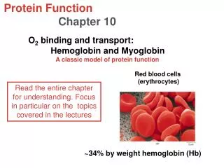
Reversible binding of a protein to a ligand: Oxygen-binding proteins Fig. 7-1,2 Heme protoporphyrin
Fig. 7-3 The structure of myoglobulin *a single binding site for O2 *78% a helices (8) *His93 or HisF8 (the 8th residue in a helix F) binds to heme *Bends between a helices
Protein-ligand interactions can be described quantitatively P + L PL Ka = [PL]/[P][L] Kd = [P][L]/[PL] q = (binding sites occupied)/(total binding sites) = [PL]/[PL] + [P] = [L]/([L] +1/Ka) q = 0.5 [L] = 1/Ka, or Kd Fig. 7-4 Graphical representation of ligand binding
P + L PL Ka = [PL]/[P][L] Dissociation constant, Kd = [P][L]/[PL] q = (binding sites occupied)/(total binding sites) = [PL]/[PL] + [P] = [L]/([L] +1/Ka) = [L]/([L] +Kd) When [L] = Kd q = 0.5 (half saturation) [L] = 9 Kd q = 0.9 Kd: the molar concentration of ligand at which half of the available ligand-binding sites are occupied Kd , affinity ( ? )
P50 = 0.26 kPa When O2 binds to Mb q = [L]/([L] +Kd) = [O2]/([O2] + Kd) = [O2]/([O2] + [O2]0.5) The concentration of a volatile substance in solution is always proportional to its partial pressure in the gas phase above the solution q = pO2/(pO2 + P50)
Protein structure affects how ligands bind 1. Steric effects 2. Molecular motions/breathing in the structure 1: 20,000 1: 200
P50 = 0.26 kPa Oxygen is transported in blood by hemoglobin (Hb) In arterial blood, Hb ~96% saturated In venous blood, Hb ~64% saturated Mb has only one subunit, as an oxygen-storage protein Mb
Fig. 7-7Comparison of aa between whale Mb and Hba, b A-H helices Only 27 aa identical
Fig. 7-8Dominant interactions between Hb subunits >30 aa 19 aa (hydrophobic, H-bonds, affected strongly upon O2 binding)
Hb undergoes a structural change on binding oxygen Fig. 7-10The T(tense) R(relaxed) transition
Fig. 7-9Some ion pairs that stabilize the T state of deoxyHb
Hb binds oxygen cooperatively 4 vs. 13.3 kPa Mb – a single subunit protein Hb – 4 subunits, an allosteric protein Fig. 7-12A sigmoid (cooperative) binding curve
Allosteric protein – a protein in which the binding of a ligand to one site affects the binding properties of another site on the same protein allos --- other stereos --- solid or shape Homotropic interaction --- liagnd = modulator Heterotropic interaction --- ligand = modulator O2 --- as both a normal ligand and an activating homotropic modulator for Hb
Cooperative ligand binding can be described quantitatively P + nL PLn Ka = [PLn]/[P][L]n Dissociation constant, Kd = [P][L] n/[PLn] Fig. 7-13 q = (binding sites occupied)/(total binding sites) = [L]n/([L]n +Kd) q /(1- q) = [L]n/Kd Log{q /(1- q)} = n log [L] – log Kd (Hill equation, 1910) Log{q /(1- q)} = n log pO2 – log P50 nH – the Hill coefficient (slope of Hill plot) <, =, > 1
Two models suggest mechanisms for cooperative binding Fig. 7-14 Concerted (all-or-none), 1965 Sequential, 1966
O2 binding to Hb is regulated by 2,3-bisphosphoglycerate (BPG) HbBPG + O2 HbO2 +BPG 4 1 [BPG] during hypoxia Fig. 7-16
++ T O2 T state BPG is negatively charged R Fig. 7-17 Binding of BPG to deoxyHb
Sickle-cell anemia is a molecular disease of Hb Val6 mutates to Glu6 in two b chains
Complementary interactions between proteins and ligands: The immune system and immunoglobulins MHC (major histocompatibility complex) all vertebrate cells macrophages, B cells
Structure of a human class I MHC protein Recognized by T-cell receptor
Induced fit in the binding of an antigen to IgG Heavy chain Light chain Kd~10-10M
The Ab-Ag interaction is the basis fro a variety of important analytical procedures Ployclonal vs. monoclonal Ab ELISA (enzyme-linked immunosorbent assay)
Protein interactions modulated by chemical energy Actin, myosin, and molecular motors Fig. 7-29 Myosin S1
The major components of muscle Fig. 7-29
Structure of skeletal muscle relaxed contracted
Molecular mechanism of muscle contraction 3~4 pN of forces, 5~10 nm movement/cycle