Chapter 5 Protein Function
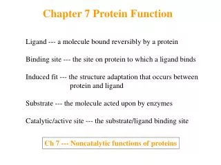
Chapter 5 Protein Function. Interaction of Proteins with Other Molecules. Ligand A molecule binding reversibly to a protein Other proteins, or any kind of molecules Binding site for a ligand Complementary to the ligand in size, shape, charge, and hydrophobic/~philic properties

Chapter 5 Protein Function
E N D
Presentation Transcript
Chapter 5 Protein Function
Interaction of Proteins with Other Molecules • Ligand • A molecule binding reversibly to a protein • Other proteins, or any kind of molecules • Binding site for a ligand • Complementary to the ligand in size, shape, charge, and hydrophobic/~philic properties • Specific & selective to one or a few ligands • Conformational change of proteins • Subtle change (breathing) • Molecular vibrations, small movement of a.a. residues • Dramatic change • Movement of major segment of a protein • Induced fit • Structural adaptation permitting tighter binding • Conformational signal • Cooperativity between ligand and protein interactions
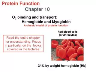
5.1 Reversible Binding of a Protein to a Ligand: Oxygen-Binding Proteins
Heme • Prosthetic group of oxygen-transporting proteins • Myoglobin, hemoglobin, cytochromes • Complex organic ring structure; Protoporphyrin • Protoporphyrin with Fe2+ (ferrous state) 6 coordination bonds for Fe2+ • 4 N in porphyrin ring • Electron donating character: prevent oxidation of Fe2+ to Fe3+ (ferric state) • 2 perpendicular to the prophyrin • 1 occupied with proximal His residue • 1 binding site for oxygen • Fe2+ ; oxygen binding • Changing from dark purple to bright red color • Higher affinity to CO and NO
Heme 4 Pyrrole rings Porphyrins
Myoglobin (Mb) • Roles of myoglobin • Oxygen transport in muscle • Abundant in diving mammals; seals and whales • Structure • 153 a.a. protein belongs to globin family • 8 a helical segments • 1 heme molecule
Protein-Ligand Interactions : Kd • Binding of O2 to myoglobin • q =[O2] / ([O2] + Kd) = [O2] / ([O2] +[O2]0.5) = [O2] / ([O2] +P50 ) P50 : local partial pressure of O2 at [O2]0.5
Protein Structure Affects How Ligands Bind • O2 and CO binding to heme • Binding to free heme • CO has more than 20,000 times higher affinity than O2 • Binding to heme in myoglobin • CO has 200 times higher affinity than O2 • Steric hindrance restricts CO binding • Roles of breathing • Heme is deeply buried inside of the protein • Rotation of distal His (10-9 sec) provides cavities for O2 entrance
Hemoglobin • Red blood cells • Generated form hemocytoblast stem cells • Hemoglobin production & carrying • Loss of intracellular organelles • Life time 120 days • Hemoglobin • In arterial blood: 96% are saturated with O2 • In venous blood: 64% are saturated with O2 • Very sensitive to O2 concentration • Good for O2 transport • Myoglobin • Relatively insensitive to O2 concentration • Good for O2 storage
Hemoglobin • Structure • 2 a (141 a.a.), 2 b (146 a.a.) chains, and 4 heme groups • Globin family of proteins • a, b chains and myoglobin • Low sequence similarity but high structural similarity • Strong interactions between a and b chains • >30 residues are involved • Mostly hydrophobic interactions
Structural Change of Hemoglobin upon Oxygen Binding • T (tense) state : low affinity O2 binding • Deoxyhemoglobin • More ion pairs at a1b2 (a2b1) interface • Slightly puckered porphyrin • R (relaxed) state : high affinity O2 binding • O2 binding state • Planar porphyrin
Cooperative Binding of Oxygen to Hemoglobin • Roles of hemoglobin • In the lung (pO2 = 13.3 kPa) : binding to O2 • In the tissues (pO2 = 4 kPa) : releasing O2 • Cooperative binding of O2 to hemoglobin • Transition form T state to R state upon O2 binding induction of conformational change of the adjacent subunit to R state • Sigmoid binding curve
Allosteric Protein • Allosteric protein • Binding of a ligand to one site affects the binding properties of another site on the same protein • Modulator : activator or inhibitor • Homotropic • Modulator = the normal ligand • Heterotropic • Modulator ≠ the normal ligand • Cooperative binding (hemoglobin) • Allosteric binding in multimeric proteins • Sigmoid binding curve • Sensitive to ligand concentration • Binding site in stable segment next to unstable segment
Quantitative Description of Cooperative Ligand Binding • Hill plot;Log (q/1-q) vs. log [L] • Slope (nH, Hill coefficient) • Degree of cooperativity • nH = 1 : no cooperativity • nH >1 : positive cooperativity • nH = n : theoretical upper limit, Simultaneous binding of the entire binding sites
Hill Plot for O2 Binding to Myoglobin and Hemoglobin • Log (q/1-q) = nlog [L] – log Kd • Log (q/1-q) = nlog pO2 – nlog P50
Models for Cooperative Binding • MWC model (concerted model) • Jaques Monod, Jeffries Wyman, Jean-Pierre Changeux (1965) • All proteins in the same conformation • Transition to high affinity conformation upon ligand binding • Sequential model • Daniel Koshland (1966) • Ligand binding induces conformational change in an individual subunit • Induce a similar change in an adjacent subunit
Transport of H+ and CO2 by Hemoglobin • Transport H+ and CO2 from the tissue to the lungs and kidneys • Carbonic anhydrase in erythrocyte • Hydration of CO2 to form bicarbonate • CO2 + H2O H+ + HCO3- • Bohr effect (1904) • Effect of [CO2]and [H+] on binding & releasing of O2 binding by hemoglobin • H+ binding : His146 in b subunit and other a.a residues stabilization of T state • HHb+ + O2 HbO2 + H+ • CO2 binding : Forms carbamate group by binding to N terminal amino group • Generation of H+ • Stabilization of T state by salt bridge
Oxygen Binding to Hemoglobin is Regulated by BPG • 2,3 bisphosphoglcerate (BPG) • Abundant in erythrocyte • Heterotropic allosteric modulator • Binding to cavity between b subunits in the T state • Interaction with positive a.a, stabilizing T state,1 BPG/Hb tetramer • Reduced O2 binding affinity of hemoglobin HbBPG + O2 HbO2 + BPG (inverse relation) • Fetal hemoglobin • a2g2 • g subunits have lower affinity for BPG • High affinity to O2 • Effective extraction of O2 from its mother’s blood
Oxygen Binding to Hemoglobin is Regulated by BPG • Facilitate O2 release in the tissue under low pO2 (high altitudes, hypoxia) • Fetal hemoglobin • a2g2 • g subunits have lower affinity for BPG • High affinity to O2 • Effective extraction of O2 from its mother’s blood
Sickle-Cell Anemia • Hemoglobin S • Glu 6 to Val mutation in two b chain (homozygote) • Heterozygote has a mild symptom • Aggregation of deoxygenated hemoglobins by hydrophobic interactions fiber formation
Sickle-Cell Anemia • Sickle shaped erythrocytes • Fragile : lower hemoglobin content • Blocking capillaries
5.2 Complementary interaction; The immune system and immunoglobulins
Immune cells • Leukocytes (white blood cells) • Recognition & binding to molecules for infection signals
Immune responses • Humoral immune system • Bacteria or virus infections • Antibodies (immunoglobulins; Ig) mediation • Binding to bacteria, viruses, other foreign molecules destruction • Produced from B lymphocytes (B cells) • Cellular immune system • Removal of infected cells & parasites/foreign tissues • T lymphocytes; cytotoxic T cells (killer T cells) • T-cell receptor-mediated recognition of infected cells or parasites • Helper T cells
Structural properties of antibodies • Immunoglobulin G (IgG) • Major class of antibody • 4 polypeptide chains; 2 heavy chains + 2 light chains • (noncovalent & disulfide bonds) • Y-shaped complex; Fa + Fab (antigen-binding fragments)
Structural properties of antibodies • Specificity between antigen and binding sites • Shape & location of noncovalent interactions • Conformational changes complete interactions • Kd value; ~ 10-10 M
Antibody techniques Enzyme-linked immunosorbent assay
5.3 Protein interaction modulated by chemical energy Contractile force generation in muscle by myosin and actin