Chapter 5 Protein Function
Lehninger Principles of Biochemistry, Fourth Edition, 2005. Chapter 5 Protein Function. 中央研究院 生物化學研究所 曾湘文 博士 Mar. 20, 2007. Three bonds separate sequential a carbons in a polypeptide chain.

Chapter 5 Protein Function
E N D
Presentation Transcript
Lehninger Principles of Biochemistry, Fourth Edition, 2005 Chapter 5Protein Function 中央研究院 生物化學研究所 曾湘文 博士 Mar. 20, 2007
Three bonds separate sequential a carbons in a polypeptide chain The N-Caand Ca -C bonds can rotate, with bond angles designated Φ (φ, phi) and Ψ (ψ, psi), respectively. The peptide CON bond is not free to rotate. Other single bonds in the backbone may also be rotationally hindered, depending on the size and charge of the R groups. In the conformation shown, φ and ψ are 180℃ (or - 180℃). As one looks out from the carbon, the φ and ψ angles increase as the carbonyl or amide nitrogens (respectively) rotate clockwise.
0o The planar peptide group and Ramachandran plot for L-Ala residues • The bond angles resulting from rotations at Ca for N-Ca - F (phi)and Ca-C - y (psi)are both defined as 0o when the two peptide bonds flaking that a carbon are in the same plane and positioned as shown. • Both f and y are defined as 180o when the polypeptide is in its fully extended conformation and all peptide groups are in the same plane Ramachandran plot
Key Words of Protein Function • Ligand: A molecule is bound reversibly by a protein. --- may be any kind of molecule, including another protein. • Binding site: A ligand binds (specifically) at a site on the protein– complementary to the ligand in size, shape, charge and hydrophobic or hydrophilic character. • Protein vs. Ligand; Catalytic (Active) site vs. Substrate; Antigen vs. Antibody • Induce fit: The structural adaptation that occurs between protein and ligand. The binding of a protein and ligand is often coupled to a conformational change in the protein that makes the binding site more complementary to the ligand, permitting tighter binding. • Conformational change in one subunit affect another subunit. • Interaction may be regulated, through specific interactions with one or more additional ligand.
Outlines: 5.1 Reversible Binding of a Protein to a Ligand: Oxygen-Binding Proteins 5.2 Complementary Interactions between Proteins and Ligands: The Immune System and Immunoglobulins 5.3 Protein Interactions Modulated by Chemical Energy: Actin, Myosin, and Molecular Motors
5.1 Reversible Binding of a Protein to a Ligand: Oxygen-Binding Proteins • Oxygen Can Be Bound to a Heme Prosthetic Group • Myoglobin Has a Single Binding Site for Oxygen • Protein-Ligand Interactions Can Be Described Quantitatively • Protein Structure Affects How Ligands Bind • Oxygen Is Transported in Blood by Hemoglobin • Hemoglobin Subunits Are Structurally Similar to Myoglobin
The heme group is present in myoglobin, hemoglobin, and many other proteins, designated heme proteins. consists of a complex organic ring structure, protoporphyrin IX, to which is bound an ion atom in its ferrous (Fe2+) state. Porphyrins, of which protoporphyrin IX is only one example, consist of four pyrrole rings linked by methene bridges, with substitutions at one or more of the positions denoted X. Heme (Haem)
Fe2+ has six coordinated bonds, four in the plane of and bonded to the flat porphyrin ring, and two perpendicular to the prophyrin. Reaction of oxygen at one of the two open coordination bonds of ion can result in irreversible conversion of Fe2+ to Fe3+ --- sequestering the heme deep within proteins. Electronic properties of heme ion change (dark purple to bright red). CO and NO has greater affinity than does O2. Two representations of Heme
The two coordination bonds to Fe2+perpendicular to the porphyrin ring system. One of these two bonds is occupied by a His93 residue (Proximal His: His-F8). The other is the binding site for oxygen. The remaining four coordination bonds are in the plane of, and bonded to, the flat porphyrin ring system. The heme group and iron
A single polypeptide of 153 AA residues with one heme. Globins family 8 a-helical segments (A to H, 78% of total AA) His (His93 or His F8) residue coordinated to the heme. The bends are designated AB, CD, EF, FG. The structure of myoglobin
Protein-Ligand interaction: equilibrium expression Kd (dissociation constant) = 1/Ka Ka: association constant
Ka:association constant; Kd:dissociation constant When [L] = Kd– half of the binding site are occupied, [L]<Kd—little ligand binds to protein. => In order for 90% of ligand-binding site to be occupied, [L] must be nine times greater than Kd. The more tightly a protein binds a ligand, the lower the concentration of ligand required for half the binding sites to be occupied, and thus, the lower the values of Kd Graphical representations of ligand binding
Steric effects on the binding of ligands to the heme of myoglobin distalHis proximal His
Steric effects on the binding of ligands to the heme of myoglobin distalHis proximal His
Protein structure affects how ligands bind • CO binds (Fe, C, O atoms in straight line) to free heme molecule over 20,000 times better than does O2 (at an angle), but only 200 times better when the heme is bound in myoglobin.--- steric hindrance. • Distal His 64 (His E7) interact molecular bound to heme.— preclude the linear binding of CO. • The binding of O2 to the heme depends on molecular motions (breathing). Heme is deeply buried in the folded polypeptide, with no direct path for oxygen to go from the surrounding solution to the ligand - binding site. Rapid molecular flexing of the aa side chain (His 64) produces transient cavities in the protein structure.
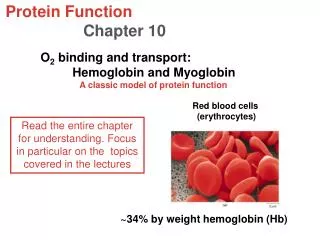
Hemoglobin subunits are structurally similar to myoblobin • Hemocytoblasts– erythrocytes (red blood cells): biconcave disks, with lots of hemoglobin (34 %) and lost nucleus, mitochondria, and endoplasmic reticulum (120 days). • Myoglobin (Mb) insensitive to O2 change => storageHemoglobin (Hb)---sensitive toO2 change => transport • Hb: tetrameric protein (a2b2). Mb Hb
The AA sequences of whale myoglobin and the a and b chains of human hemoglobin • Hb and Mb (27 identical)--- 3-D structure are similar. • a subunit lacks the short D helix. The heme-binding pocket is made up largely of the E and F helices.
a1b1 Interface (anda2b2 counterpart)– 30 AA involved. And is sufficiently strong that mild treatment of Hb with urea tends to cause the tetramer to disassembles into ab dimers => a1b1 +a2b2 . a1b2 (and a2b1) interface involved 19 AA. Hydrophobic interactions predominated, also H bonds and ion pairs (salt bridges). Dominant interactions between Hb subunits 30 aa 19 aa
T state: O2 absent. R state: stabilized in O2 binding. Interaction between the ion pairs His HC3 and AspFG1 of the b subunit and between Lys C5 of the a subunit and the a-carboxyl group of His HC3 of the b subunit. Some ion pairs that stabilized the T state of dexoyhemoglobin
The T to R transition: Hb undergoes a structural change on binding O2 narrowing the pocking between the b subunits • T state (Tense): Hb is stabilized by a greater number of ion pairs, which lie at the a1b2 (and a2b1) interface Vs. R state (Relaxed): O2 has higher affinity. • The binding of O2 to a Hb subunit in the T state triggers a change in conformation to the R state --- individual subunits change little, but ab subunit pairs slide past each other and rotate, narrowing the pocking between the b subunits --- old ion pairs broken but new ones are formed.
The shift in the position of the F helix when heme binds O2 is one of the adjustment that is believed to trigger the T to R transition. In the T state– porphyrin is slightly puckered 皺起 -- heme to protrude somewhat on the proximal His (His F8) side; Binding of O2 cause heme to assume amore planar conformation, shifting the proximal His and the attached F helix. Val in the E helix (Val E11) blocks the heme in the T state –swing out of the way for O2 to bind--- adjustments of the ion pairs at the a1b2 interface. Change in conformation near heme on O2 binding puckered more planar
A sigmoid (cooperative) binding curve: Hb binds O2 cooperatively • Sigmoid binding curve– a transition from a low-affinity to a high-affinity state. • A single-subunit protein with a single ligand-binding site cannot produce a sigmoid bindinb curve • Cooperative binding renders Hb more sensitive to the small differences in O2 concentration between the tissues and the lungs, allowing Hb to bind O2 in the lung where O2 is high and release it in the tissues where pO2 is low. • Allosteric protein (other shape) : protein in which the binding of a ligand (modulators) to one site affects the binding properties of another site on the same protein. • Homotropic allosteric protein (Hb and O2) Vs. heterotropic.
Hill plots for the binding of oxygen to myoglobin and hemoglobin • Based on the equation: slope= n (nH, Hill coefficient). The experimentally determined slope ( the degree of interaction, not the number of binding sites). • nH>1, ligand binding cooperatively; nH=1 not cooperative. nH<1, negative cooperatively. • Upper limit nH = n
Hill plots for the binding of oxygen to myoglobin and hemoglobin • When nH =1, there is no evident cooperativity. • The maximum degree of cooperativity observed for hemoglobin corresponds approximately to nH = 3. • Note that while this indicates a high level of cooperativity, nH is less than n, the number of O2-binding sites in hemoglobin. • This is normal for a protein that exhibits allosteric binding behavior.
Mechanisms for cooperative binding: Two models • MWC (concerted) model (all-or-none): assumes that the subunits of a cooperatively binding protein are functionally identical, that each subunit can exist in (at least) two conformations, and that all subunits undergo the transition from one conformation to the other simultaneously (no protein has individual subunits in different conformation) two conformations are in equilibrium, Substrate can bind both conformations (different affinity). • Sequential model: Ligand binding can induce a change of conformation in an individual subunit. A conformational change in one subunit makes a similar change in an adjacent subunit (making binding of secondary subunit more likely MWC (concerted) model Sequential model
Effect of pH on the binding of oxygen to Hemoglobin • Bohr effect: The effect of pH and CO2 concentration on the binding and release of oxygen by hemoglobin. • Carbonic anhydrase (production of H+), the binging of H+, CO2 inversely related to the binding of O2. When O2 is high (lung), Hb binds O2 and release H+. When O2 is low (peripheral tissues), Hb binds H+, and release O2. • H+ bind His146 of b subunits--- ion pairs to Asp94—T state. • CO2 binds as a carbamated group to the a-amino group at amion-terminal end of each globin chain---salt bridge.
Effect of BPG on the binding of Oxygen to Hb • Oxygen binding to hemoglobin is regulated by 2,3-Bisphosphoglycerate • In RBC (erythrocytes)---reduce the affinity of Hb for O2. Binds to Hb in the cavity between the b subunits in the T state (negative charges of BPG interact with several positively charged group). Only one BPG is bound to each Hb tetramer. • Adjustment in the BPG level (5 mM to 8 mM) has a small effect on the binding of O2 in the lungs but a considerable effect on the release of O2 in the tissues. • Hypoxia: lowered oxygenation of peripheral tissues due to inadequate function of the lungs or circulatory system.
Binding of BPG to deoxyhemoglobin • BPG binding stabilizes the T state of deoxyhemoglobin, shown here as a mesh surface image. • The negative charges of BPG interact with several positively charged groups (blue) that surround the pocket between the b subunits in the T state. • The binding pocket for BPG disappears on oxygenation, following transition to the R state. • Fetuses, g subunits (a2g2) Hb has much lower affinity for BPG than normal adult hemoglobin, and a correspondingly higher affinity for O2.
Sickle-cell anemia is a molecular disease of Hemoglobin • A comparison of uniform, cup-shaped, normal erythrocytes (a) with the variably shaped erythrocytes seen in sickle-cell anemia (b), which range from normal to spiny or sickle-shaped. • The sickle-cell anemia patient suffer from repeated crises brought on by physical exertion (weak, dizzy, short of breath, and heart murmurs and an increased heart rate): has few and abnormal erythrocytes, lots of immature cell, the blood contains many long, thin, crescent-shaped erythrocytes (blade of sickle)---insoluble and forms polymers that aggregate into tubular fiber (HbS) • HbS: a Glu to Val substitution at position 6 in 2 b chains. Has fewer negative charge– creates a sticky hydrophobic contact point at outer surface of Hb--- associate abnormally with each other--- long, fibrous aggregate.
Normal and sickle-cell hemoglobin • Subtle differences between the conformations of hemoglobin A and hemoglobin S result from a single amino acid change in the chains. • As a result of this change, deoxyhemoglobin S has a hydrophobic patch on its surface, which causes the molecules to aggregate into strands that align into insoluble fibers.
5.2 Complementary Interactions between Proteins and Ligands: The Immune System and Immunoglobulins • Immunity is brought about by a variety of leukocytes (white blood cells), including macrophages and lymphocytes. • The immune response consists of two complementary systems. • The humoral immune system (Latin humor, “fluid”) is directed at bacterial infections and extracellular viruses (those found in the body fluids), but can also respond to individual proteins introduced into the organism. • The cellular immune system destroys host cells infected by viruses and also destroys some parasites and foreign tissues.
Complementary interactions between proteins and ligand: The immune system and immunoglobulins • Humoral (fluid) immune system: directed at bacterial infections and extracellular viruses--- antibodies or immunoglobulins (Ig)--- produced and secreted by B cells. • Cellular immune system: destroys host cells infected by viruses and parasites and foreign tissues.---T cells. • Antigen: any molecule or pathogen capable of eliciting an immune response. --- complex antigen may be bound by a number of different Abs within the antigen– antigenic determinant (epitope). • Haptens: Non-antigenic small molecules (<5,000) that linked to larger proteins may elicit an immune response. Abs produced in response to protein-linked haptens will then bind to the same small molecules when they are free.
MHC (Major Histocompatibility Complex) proteins • These proteins consist of a and b chains. • In class I MHC proteins, the small b chain is invariant but the amino acid sequence of the a chain exhibits a high degree of variability, localized in specific domains of the protein that appear on the outside of the cell. • Each human produces up to six different a chains for class I MHC proteins. • In class II MHC proteins, both the a and b chains have regions of relatively high variability near their amino-terminal ends.
MHC (Major Histocompatibility Complex) proteins • Class I (all vertebrate cells): consist of a and b chains, the small b chain is invariant but the amino acid sequence of the a chain exhibits a high degree of variability, localized in specific domains of the protein that appear on the outside of the cell. Each human produces up to six different a chains for class I MHC proteins--- highly polymorphic • Binds and display peptides derived from the proteolytic degradation and turnover of proteins that occurs randomly within the cell—recognition targets of the T-cell receptors of the Tc cells in the cellular immune system. • Each Tc cell has many copies of only one T-cell receptor that is specific for a particular class I MHC • The maturation of Tc cells in the thymus includes a stringent selection process that eliminates more than 95% of the developing Tc cells, including those that might recognize and bind class I MHC proteins displaying peptides from cellular proteins of the organism itself. The Tc cells that survive and mature are those with T-cell receptors that do not binds to organism’s own proteins • Class II (on the surface of macrophages and B lymphocytes): both the a and b chains have regions of relatively high variability near their N terminal ends. • Binds and display peptides derived not from cellular proteins but from external proteins but from external proteins ingested by the cells--- binding targets of the TH cell receptors
Structure of a human class I MHC protein • This model is derived in part from the known structure of the extracellular portion of the protein (PDB ID 1DDH). The chain of MHC is shown in gray; the small chain is blue; the disulfide bonds are yellow. A bound ligand, a peptide derived from HIV, is shown in red. • Top view of the protein, showing a surface contour image of the site where peptides are bound and displayed. The HIV peptide (red) occupies the site. This part of the class I MHC protein interacts with T-cell receptors.
Immunoglobulins (Igs) • 5 classes of Igs (by heavy chains): IgA-a, IgD-d, IgE-e, IgG-g, IgM-m. light chains (k, l). • IgA(mono-, di-, trimer): found in secretions, saliva, tears, milk. Is the first Ab made by B cells and is primary Ab in early immune response. • IgD (monomer): has same antigen binding site as IgM by the same B cells. • IgE(monomer): plays an important role in the allergic response, and interacts with basophils (phagocytic leukocytes) in blood and histamine-secretion (mast) cells in tissue. IgE binds to Fc receptor on basophils to secretion of histamine- dilation and increased permeability of blood vessel (movement of Immune response cells and Igs to inflammation site).. • IgG (monomer): the major Ab in secondary immune responses (initiated by memory B cells. Most abundant Igs in blood. Binds to invading bacterium or virus directly and activated the complement system, activating macrophages to engulf and destroy the invader. • IgM (monomer—membrane-bound, pentamer– secreted)
The structure of immunoglobulin G (IgG) • Pairs of heavy and light chains combine to form a Y-shaped molecule. Two antigen-binding sites are formed by the combination of variable domains from one light (VL) and one heavy (VH) chain. Cleavage with papain separates the Fab (antigen-binding) and Fc (readily crystallizes) in the hinge region. The Fc regions– have carbohydrate. • Three constant domain in each heavy chains, and one in light chain. Both have one variable domain each. The variable domains associated to create the antigen-binding site. Conformational flexibility may be important to the function of immunoglobulins.
Binding of IgG to and antigen • To generate an optimal fit for the antigen, the binding sites of IgG often undergo slight conformational changes. Such induced fit is common to many protein-ligand interactions.
Binding of IgG to and antigen • To generate an optimal fit for the antigen, the binding sites of IgG often undergo slight conformational changes, such induced fit is common to many protein-ligand interactions. • The binding cavity has enlarged and several groups have shifted position (IgG and HIV Ag)
IgM pentamer of immunoglobulin units • IgM(monomer—membrane-bound, pentamer– secreted): • The pentamer is cross-linked with disulfide bonds (yellow). • The J chain is a polypeptide of Mr 20,000 found in both IgA and IgM.
Phagocytosis of an antibody-bound virus by a macrophage • The Fc regions of the antibodies bind to Fc receptor on the surface of the macrophage, triggering the macrophage to engulf and destroy the virus---phagocytosis.
The antibody-antigen interaction is the basis for a variety of important analytical procedures • Polyclonal antibodies: are those produce by many different B lymphocytes responding to one antigen– Abs bind different epitopes within the Ags. • Monoclonal antibodies, are produce by a population of identical B cells (a clone) grown in cell culture. Abs are homogeneous, recognizing the same epitope. • Usage of Ab: Affinity chromatography, Gene inactivation, ELSA…
ELSA - Enzyme-Linked ImmunoSorbentAssay • For rapid screening and quantification of the presence of an Ag in a sample. • Wells are coated with Ag.—block proteins with inexpensive non specific protein (milk)---First Ab—wash---secondary Ab with an enzyme—wash--- substrate for the Ab linked enzyme (color)
Immunoblot assay - Western blot • Proteins are separate by SDS-PAGE and transfer to nitrocellulose membrane • Block • primary Ab • secondary Ab linked with enzyme • substrate for enzyme • detection of minor component in sample with M.W.
5.3 Protein Interactions Modulated by Chemical Energy: Actin, Myosin, and Molecular Motors
Protein interactions modulated by chemical Energy:Actin, Myosin, and molecular motors • Myosin (2 heavy chains + 4 light chains): C-terminals are arranged as extended a helices – left handed, similar to a-keratin. N-terminals (globular domain)—ATP hydrolyzed. • Treat with trypsin: light and heavy meromyosin. • Papain: myosin subfragment 1 (S1), myosin head group
The major components of muscle rodlike- long bipolar • Thick filaments: myosin aggregated—rodlike structure. Several hundred molecules arranged with their fibrous tails, long bipolar structure. • Thin filament: F-actin, troponin (TN) and tropomysin (TM) -- assembled as successive monomeric actin molecules add to one end.
Structure of Skeletal muscle • Bundles of muscle fibers— a single, very large, multinucleated cell --- 1000 myofibrils (regularly arrayed thick and thin filaments complex. • Sarcoplasmic reticulum (flat membranous vesicles) surround each myofibril.