Understanding X-ray Interactions in Matter
290 likes | 455 Vues
Explore the intricate interactions between X-rays and matter through coherent scattering, Compton scattering, and the photoelectric effect. Learn how different tissues absorb radiation and the role of secondary electrons. Discover the significance of beam attenuation and radiation dose measurements.

Understanding X-ray Interactions in Matter
E N D
Presentation Transcript
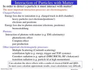
INTERACTION OF X RAYS WITH MATTER
INTERACTION BETWEEN X-RAYS AND MATTER Coherent Scattering Photoelectric effect Compton Scattering
COHERENT SCATTERING 8 % Classic scattering Incident X-ray photon Scattered X-ray photon Low energy photons only scattering, no Ionization and only little energy loss
COHERENT SCATTERING – Rayleigh (coherent) scatter interaction with entire atom . Photon changes direction (though usually by small angle) . “Elastic” scattering: Photon energy unchanged . Most important for “soft” (low energy) . and x rays (< 10 keV) . Adds very little to film fog
COMPTON SCATTERING Compton effect or Compton scattering (C) incoherent scattering: Directly proportional to electron density Can occur with very low atomic weight targets even at relatively low X-ray energies 62 %
COMPTON SCATTERING • An x-ray photon of lower energy is scattered from the • atom • Proportional to electron density of absorber • Interaction with outer electron • Changes direction • Adds to film fog 62 %
COMPTON SCATTERING • Incident X-ray changes direction and loses energy, imparting that energy to the electron (Compton electron) • Compton electron interacts with other atoms producing secondary ionizations • Possess relatively low energy, produce low energy x- rays • Energies of 100 keV -- 10 MeV – absorption of radiation mainly due to the Compton effect
PHOTOELECTRIC EFFECT Interaction with bound atomic electron . Incident photon disappears . Photon energy absorbed by electron . Momentum absorbed by atom . Probability of PE absorption increases for: » Low incident photon energy » High electron density in medium (mass density x Z) . Following absorption, photoelectrons, and “characteristic” x rays are emitted
PHOTOELECTRIC EFFECT 30 % • Critical indiagnostic X rays • Ionization occurs • Binding energy is small for biologic tissues • Usually with - K shell electron • Recoil electron travels only short distance
DIFFERENTIAL ABSORPTION Different tissues - different amount of absorption Makes radiographs possible Hard tissue - mostly absorbed Soft tissue - heterogeneous beam Mainly by photoelectric effect - varies with third power of Z Of absorber 6.5 times more in bone than water
SECONDARY ELECTRONS • Electrons ejected by photoelectric effect or • Compton effect • Energy given up by: • Collisional ionization or excitation • Radiative Bremsstrahlung radiation low energy
BEAM ATTENUATION Reduction in intensity while passing through mattter Either by photo electric effect or Compton scattering Half Value Layer - thickness of an absorber needed to reduce the number of x ray photon by half
RADIATION DOSE The International System (SI) unit • Radioactivity • Exposure • Absorbed Dose • Dose Equivalent • Effective dose
RADIATION DOSE Activity: rate at which the isotope decays. Radioactivity may be thought of as the volume of radiation produced in a given amount of time. International System (SI) unit - Becquerel (Bq) (curie (Ci) Exposure: measure of the strength of a radiation field at some point in air. measure made by a survey meter. Gray Gy (Roentgen (R) Effective dose: Sum of product of equivqlent dose to each organ or tissue Sivert Si
RADIATION DOSE Absorbed Dose:amount of energy that ionizing radiation imparts to a given mass of matter. SI unit gray (Gy), “rad” (Radiation Absorbed Dose) 1 rad - 0.01 Gy Different materials that receive the same exposure may absorb different energy Dose Equivalent: relates the absorbed dose to the biological effect of that dose absorbed dose of specific radiation X "quality factor" = dose equivalent (SI unit - sievert (SV), (rem)" One rem = 0.01 SV X- or Gamma radiation - the quality factor is 1
DOSIMETRY Quantity of radiation exposure Radiation dosimeters are devices which are capable of measuring an accumulated absorbed dose of ionizing radiation • issued four times a year • - rate measuring instruments and - personal dose measuring instruments • Direct • Indirect.
DOSIMETERS Film Badges Ring Dosimeters
DOSIMETERS Pocket Dosimeter - Direct Read Pocket Dosimeter Digital Electronic Dosimeter Ionization Counters Scintillation Counters Particle Track Devices The Cloud Chamber Geiger Muller Counter
THERMOLUMINESCENT DOSIMETERS (TLDS) 15% for low doses 3% for high doses TLD - a phosphor, such as lithium fluoride (LiF) or calcium fluoride (CaF), in a solid crystal structure exposed to ionizing radiationradiation interaction ionization trapping of free electrons Heating the crystal Release of energy as light 1 millirem
Thermoluminescence dosimeter T L material T L D reader EXPOSURE Equipment used to measure T L heated Emitted light proportonal to absorbed dose
Parts of TLD reader • HEATER • PHOTOMULTIPLIER TUBE • HEAT FILTER ALOW THE LIGHT TO PASS • MEASURE THE EMMITED LIGHT • ELECTRONIC SYSTEM • (POWER SUPPLY)
AVAILABILITY • TLD AVAILABLE IN • SOLID • POWDER • APPLICATION OF TLD • RADIOTHERAPY • RADIODIAGNOSIS • PERSONAL MONITORING
P M TUBE FILTER TLD READER PROCEDURE TRAY HEATER