Download
1 / 26
270 likes | 606 Vues
6.1 Ectodermal competence and the ability to respond to the optic vesicle inducer in Xenopus. Cell Communication

E N D
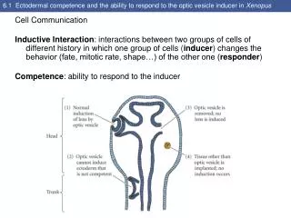
6.1 Ectodermal competence and the ability to respond to the optic vesicle inducer in Xenopus Cell Communication Inductive Interaction: interactions between two groups of cells of different history in which one group of cells (inducer) changes the behavior (fate, mitotic rate, shape…) of the other one (responder) Competence: ability to respond to the inducer
6.2 Induction of optic and nasal structures by Pax6 in the rat embryo Competence is not a passive state but an actively acquired condition. Pax6 protein appears to be important in making the ectoderm competent to respond to the inductive signal from the optic vesicle.
6.3 Recombination experiments show that the induction deficiency of Pax6-deficient rats is caused by the inability of the surface ectoderm to respond to the optic vesicle
6.5 Schematic diagram of the induction of the mouse lens (Part 1) • At E9, the optic vesicle extends toward the surface ectoderm. • At E9.5, the lens placode has enlarged and the optical vesicle has formed an optic cup. • At E10.5, the central porting of the lens forming ectoderm invaginates, while the 2 layers of retina become distinguished.
6.5 Schematic diagram of the induction of the mouse lens (Part 2) D) At E11.5, the lens vesicle has formed. E) At E13, the lens consists of anterior cuboidal epithelial cells and elongating posterior fiber cells. The cornea develops in front of the lens.
6.5 Schematic diagram of the induction of the mouse lens (Part 3) Summary of some of the inductive interactions during eye development.
Instructive and permissive interactions • Instructive interaction: a signal from an inducing cell is necessary for initiating new gene expression in the responding cell.Example: optic vesicle placed in a new region of the head ectoderm • Permissive interaction: the responding tissue has already been specified, and needs only an environment that allows the expression of these traits. Example: fibronectin and laminin as a solid substrate
6.6 Feather induction in the chick All organs consist of an epithelium and an associated mesenchyme. Epithelia- sheets or tubes of connected cells; can originate from any germ layer. Mesenchyme- loosely packed, unconnected cells; derived from mesoderm or neural crest. E10Sonic hedgehog expression
6.7 Regional specificity of induction in the chick Specific cutaneous structures can be the broad feathers of the wing, the narrow feathers of the thigh, or the scales and claws of the feet. Dermal mesenchyme is responsible for the regional specificity of induction in the competent epidermal epithelial.
6.8 Genetic specificity of induction in amphibians The response of the epithelial is species-specific.
6.9 Mechanisms of inductive interaction Juxtacrine interactions: when cell membranes proteins on one cell surface interact with receptor proteins on adjacent cells. Paracrine interactions: when proteins synthesized by one cell can diffuse over small distances to induce changes in neighboring cells (paracrine factors or GDFs). Endocrine factors: travel through the blood to exert their effects. Autocrine regulation: the same cell that secretes paracrine factors also respond to them.
6.11 Fgf8 in the developing chick • Distal limb bud ectoderm • Somitic mesoderm • Boundary between the hindbrain and midbraind • Developing eye • Tail
6.13 Activation of the Mitf transcription factor through the binding of stem cell factor by the Kit RTK protein (Part 1)
6.13 Activation of the Mitf transcription factor through the binding of stem cell factor by the Kit RTK protein (Part 2)
6.14 A STAT pathway: the casein gene activation pathway activated by prolactin
6.15 A mutation in the gene for FgfR3 causes the premature constitutive activation of the STAT pathway and the production of phosphorylated Stat1 protein
6.16 The sonic hedgehog gene is shown by in situ hybridization to be expressed in the chick nervous system (red arrow), gut (blue arrow), and limb bud (black arrow) of a 3-day chick embryo
6.18 Head of a cyclopic lamb born of a ewe who had eaten Veratrum californicum early in pregnancy The cerebral hemispheres are fused, forming only one central eye and no pituitary gland.
6.19 Wnt proteins play several roles in the development of the urogenital organs Wn4 is necessary for kidney development and for female sex determination. E14- expression is seen in the mesenchyme that condenses to form the kidney’s nephrons. Wild type and knockout Wnt4 (kidneys fail to develop)
6.20 The Wnt signal transduction pathways (Part 1) APC: adenomatosis polyposis coli TCF/LEF: T cell factor /lymphoid enhancer factor