Download
1 / 32
320 likes | 472 Vues
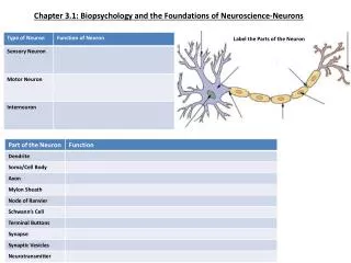
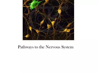
LO 2.2. The part of the neuron responsible for carrying a message from one side of a neuron to the other is called: Soma Dendrite Axon Glial cells Myelin. LO 2.2. The part of the neuron responsible for carrying a message from one side of a neuron to the other is called: Soma

E N D
LO 2.2 The part of the neuron responsible for carrying a message from one side of a neuron to the other is called: Soma Dendrite Axon Glial cells Myelin
LO 2.2 The part of the neuron responsible for carrying a message from one side of a neuron to the other is called: Soma Dendrite Axon (p. 45) Glial cells Myelin
LO 2.2 ________ is a fatty substance that forms a protective coating around the axon of a neuron. • Dendrite • Soma • Terminal branches • Neurilemma • Myelin
LO 2.2 ________ is a fatty substance that forms a protective coating around the axon of a neuron. • Dendrite • Soma • Terminal branches • Neurilemma • Myelin (p. 46)
LO 2.2 When an action potential occurs, _______ ions come into a section of the axon to make it more positive. • sodium • chloride • neurotransmitter • potassium • hydrogen
LO 2.2 When an action potential occurs, _______ ions come into a section of the axon to make it more positive. • sodium (p. 48) • chloride • neurotransmitter • potassium • hydrogen
LO 2.3 _________ are sections on a dendrite onto which neurotransmitters attach so a message can be received by a neuron: • Synapse vesicles • Synaptic gap • Receptor sites • Action potentials • Resting potentials
LO 2.3 _________ are sections on a dendrite onto which neurotransmitters attach so a message can be received by a neuron: • Synapse vesicles • Synaptic gap • Receptor sites (p. 50) • Action potentials • Resting potentials
LO 2.4 _______ is an inhibitory neurotransmitter that helps to reduce anxiety by binding to the same receptor sites that are affected by tranquilizers and alcohol. • Serotonin • GABA • Acetylcholine • Glutamate • Dopamine
LO 2.4 _______ is an inhibitory neurotransmitter that helps to reduce anxiety by binding to the same receptor sites that are affected by tranquilizers and alcohol. • Serotonin • GABA (p. 51) • Acetylcholine • Glutamate • Dopamine
LO 2.4 An excitatory neurotransmitter that is involved with memory is called: • Serotonin • GABA • Glutamate • Acetylcholine • Norepinephrine
LO 2.4 An excitatory neurotransmitter that is involved with memory is called: • Serotonin • GABA • Glutamate • Acetylcholine (p. 51) • Norepinephrine
LO 2.5 The ___________ nervous system is made up of the brain and spinal cord. • peripheral • autonomic • somatic • parasympathetic • central
LO 2.5 The ___________ nervous system is made up of the brain and spinal cord. • peripheral • autonomic • somatic • parasympathetic • central (p. 53)
LO 2.6 This section of the nervous system is responsible for calming the body after a stressful response: • Sympathetic • Central • Parasympathetic • Autonomic • Sensory neurons
LO 2.6 This section of the nervous system is responsible for calming the body after a stressful response: • Sympathetic • Central • Parasympathetic (p. 58-59) • Autonomic • Sensory neurons
LO 2.8 _________ is a technique used to study the brain that involves injecting radioactive glucose to detect activity in the brain during various tasks. • MRI scan • PET scan • CT scan • EEG • Deep lesioning
LO 2.8 _________ is a technique used to study the brain that involves injecting radioactive glucose to detect activity in the brain during various tasks. • MRI scan • PET scan (p. 62) • CT scan • EEG • Deep lesioning
LO 2.9 This section of the brain is located at the top of the spinal column and is involved with life-sustaining functions such as heart rate, respiration, and swallowing: • Pons • Reticular formation • Medulla • Thalamus • Hypothalamus
LO 2.9 This section of the brain is located at the top of the spinal column and is involved with life-sustaining functions such as heart rate, respiration, and swallowing: • Pons • Reticular formation • Medulla (p. 63) • Thalamus • Hypothalamus
LO 2.9 A patient in a hospital has difficulty controlling fine motor movement, coordinating simple movements that are involved in more complex movements (e.g., walking), and has difficulty with balance. The brain area that is most likely damaged is called: • Reticular formation • Cerebellum • Medulla • Pons • Thalamus
LO 2.9 A patient in a hospital has difficulty controlling fine motor movement, coordinating simple movements that are involved in more complex movements (e.g., walking), and has difficulty with balance. The brain area that is most likely damaged is called: • Reticular formation • Cerebellum (p. 64) • Medulla • Pons • Thalamus
LO 2.10 The __________ is involved with responses related to fear relatively quickly, allowing people to respond to danger sometimes before even being consciously aware that it exists: • amygdala • thalamus • hypothalamus • hippocampus • pons
LO 2.10 The __________ is involved with responses related to fear relatively quickly, allowing people to respond to danger sometimes before even being consciously aware that it exists: • amygdala (p. 66) • thalamus • hypothalamus • hippocampus • pons
LO 2.12 This area of the brain is responsible for higher mental functions, such as planning, personality, memory, and decision making: • Temporal lobes • Parietal lobes • Frontal lobes • Occipital lobes • Motor cortex
LO 2.12 This area of the brain is responsible for higher mental functions, such as planning, personality, memory, and decision making: • Temporal lobes • Parietal lobes • Frontal lobes (p. 68) • Occipital lobes • Motor cortex
LO 2.12 ________ is an association area located in the left temporal lobe that is responsible for understanding the meaning of language. • Broca’s area • Wernicke’s area • Somatosensory cortex • Corpus callosum • Motor cortex
LO 2.12 ________ is an association area located in the left temporal lobe that is responsible for understanding the meaning of language. • Broca’s area • Wernicke’s area (p. 69) • Somatosensory cortex • Corpus callosum • Motor cortex
LO 2.13 The area of the brain that connects the two cerebral hemispheres and is often severed in split brain patients is called: • Temporal lobe • Parietal lobe • Occipital lobe • Frontal lobe • Corpus callosum
LO 2.13 The area of the brain that connects the two cerebral hemispheres and is often severed in split brain patients is called: • Temporal lobe • Parietal lobe • Occipital lobe • Frontal lobe • Corpus callosum (p. 67)
LO 2.14 _________ is a hormone that is implicated in sleep-wake cycles: • Insulin • Melatonin • Cortisol • Glucagons • Thyroxin
LO 2.14 _________ is a hormone that is implicated in sleep-wake cycles: • Insulin • Melatonin (p. 73) • Cortisol • Glucagons • Thyroxin