Download

1 / 48
480 likes | 705 Vues
Seronegative Spondyloarthropathies. Jaya Ravindran Rheumatologist. Introduction. Cases Overview sero-ve diseases. Case 1. A 34-year-old secretary 3 months painful swelling of her right 2 nd and 4th fingers

E N D
Seronegative Spondyloarthropathies Jaya Ravindran Rheumatologist
Introduction • Cases • Overview sero-ve diseases
Case 1 • A 34-year-old secretary • 3 months painful swelling of her right 2nd and 4th fingers • 2 weeks later tenderness and swelling in the 2nd MCPs and the 3rd and 5th right PIPs, diffuse painful swelling of the 3rd toe of her left foot.
Case 2 • 22-year-old man, 3 months history of pain in 2 areas of his left foot (toes and heel). • left knee has been getting sore and stiff. • Relevant Questions?
Case 2 • 1months ago, he developed nausea, cramps, and diarrhoea after attending an "all-you-can-eat" buffet. • eyes "scratchy" of late • some burning when he urinates
Case 3 • 21-year-old male student • low back pain of 6 months' duration. • Relevant questions?
Case 3 • The onset insidious over the course of the previous 6 months. • worse in the morning, improves with activity • wakes up in the middle of the night with back pain that goes away after he walks around. • pain is located in the low back and intermittently goes down the back of one leg or the other to the knee. • He has an uncle, age 50, who has "always" had a stiff back. • painful red eye 6 months ago, which was treated by an ophthalmologist for 2 months at university.
Case 3 • Diagnosis? • Likely ocular diagnosis? • Investigations?
Investigations • XR SIJ and L/Spine normal • CRP, ESR normal
Investigations • HLA-B27 +ve - referred • MRI bilateral sacroiliitis
Spectrum • Ankylosing spondylitis • Psoriatic arthritis • Reactive arthritis • Enteropathic arthritis • Undifferentiated spondyloarthritis • Juvenile AS
Demography AS • Prevalence AS 0.05-0.23%, 3-4X male • UHCW catchment area – 375-1700 AS pts
Burden of AS • SMR 1.5 • 10% less labour participation • 15% constraints at work • Poor quality of life cf worse than RA
Aetiology • AS has been closely associated with the expression of the HLA-B27 gene • The response to the therapeutic blockade of TNFalpha indicates that this cytokine plays a central role in AS • Examination of inflamed SI joints in AS patients has demonstrated high levels of CD4+ and CD8+ T cells and macrophages. • The overlapping features with reactive arthritis and IBD (SpAs) suggests a possible role for intestinal bacteria in the pathogenesis of AS.
Diagnostic criteria – Modified New York criteria • Radiologic criteria : sacroiliitis - grade 2 bilaterally or grade 3-4 unilaterally • Clinical criteria : LBP and stiffness > 3 months improved with exercise and not relieved by rest, limitation of L/spine motion in frontal and sagittal planes, limitation of chest expansion relative to normal values correlated with age and sex • Diagnosis :radiologic criteria and at least one clinical
AS Clinical Features - axial • Early AS Romanus lesion • Advanced AS bony ankylosis
AS Clinical Features - peripheral • 30% hip and shoulder disease • Peripheral enthesopathy
Complications - Fracture • Traumatic • C5/6 also C6/7 and C7/T1 • Unstable – immobilization and fixation • Osteoporotic (20-60%) and vertebral fractures (8-15%) • Discitis
5%, dorsal spine Inflammatory Posterior # and instability Complications - Spondylodiscitis
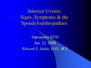
AS Clinical Features – extra-articular - Uveitis • 20-30% • B27 +ve • Acute unilateral pain, increased lacrimation, photophobia, blurred vision • Circumcorneal congestion, iris discoloured • Pupil small (irregular) • Slit lamp – exudates In anterior chamber
AS extra-articular features • Psoriasis 10-15%
AS Clinical Features – extra-articular – Inflammatory bowel • GI - Clinically silent enteric mucosal lesions 30-60% • UC and Crohn’s 5-15% spinal and 10-20% peripheral arthritis
AS Clinical Features – extra-articular - Cardiac • 2% • Increases with age, duration and peripheral arthritis • Aortic regurgitation – 3.5% (after 15years) and 10% (after 30 years) • Conduction defects – 2.7% (after 15years) and 8.5% (after 30 years)
AS Clinical Features – extra-articular - Upper lobe fibrosis • 1.3% • 20 years after onset • Bilateral linear or patchy opacities • Later cystic • Colonized by aspergillus
AS Clinical Features – extra-articular • Neurological – fracture dislocation, Cauda equina syndrome, atlanto-axial disease • Renal – amyloidosis, IgA nephropathy, analgesic nephropathy
Investigations • L/spine and SIJ x-rays • CRP and ESR • HLA B-27 – high clinical suspicion but x-ray not diagnostic – if positive worth referring as MRI can confirm pre-radiographic AS
AS – treatment • Physiotherapy • NSAIDS • ‘DMARDs’ and steroids • TNF alpha blockade • Surgery
Demography - PsA • No widely accepted criteria for diagnosis of PsA • BSR guidelines estimate prevalence of 0.1% -1% - 500-1000 patients in UHCW • Peak age of onset: 35-50 years • Equal sex distribution
Burden of PsA • 40%–57% have deforming arthritis • 11%–19% are disabled • Mortality is increased, compared with general population
PsA – clinical features 5 clinical subgroups: • (Symmetrical) polyarthritis (RA-like) – 50% cases • Asymmetrical oligoarthritis - 35% cases • DIP disease - 5% cases • Spondylitis (axial involvement) – 5% cases • Arthritis mutilans - 5% cases ……..but much overlap
Treatment • NSAIDs • DMARDs – Sulphasalazine, Methotrexate, Leflunomide, Cyclosporin • Steroids • TNF alpha blockade • OT, PT • Surgery • Dermatology input
Reactive arthritis • Young adults, equal sex • Incidence of 30-40/100,000 • Post urethritis/cervicitis or infectious diarrhoea eg campylobacter, salmonella, shigella, yersinia,chlamydia – 1-6 weeks • Sero-ve features + conjunctivitis, balanitis, oral ulcers, pustular psoriasis
Reactive arthritis • Culture – throat, urine, stool, urethra/cervix • Treatment – NSAIDs, steroids –intra-articular, antibiotics – chlamydia, DMARDs eg sulphasalazine
Summary • Young adults • Enthesitis, peripheral arthritis, spinal inflammation • Psoriasis, inflammatory bowel disease, anterior uveitis, prior GU/GI infection • B27 screening in inflammatory back pain with normal x-rays • TNF alpha blockers – new hope