Download

1 / 38
521 likes | 3.33k Vues
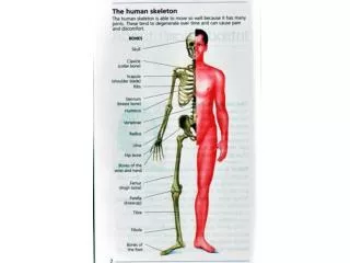
SERONEGATIVE ARTHRITIS. A group of inflammatory conditions causing arthritis of the spine and peripheral joints, often associated with HLA B27, but without a positive rheumatoid factor, hence "seronegative".

E N D
A group of inflammatory conditions causing arthritis of the spine and peripheral joints, often associated with HLA B27, but without a positive rheumatoid factor, hence "seronegative". • The joint involvement is more limited than in rheumatoid arthritis, and has different distribution - the lumbo-sacral spine, distal interphalangeal joints, depending on the subtype. • Enthesopathy (ligament/ tendon to bone junction inflammation) is more common. • Share similar pathogenesis with RA
Seronegative arthritis are more common in Males than in Females
Types: • Ankylosing spondylitis • Reiter’s syndrome • Psoriatic Arthropathy • Arthropathy associated with IBD
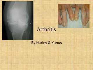
Ankylosing Spondylitis • It is a chronic, inflammatory arthritis • It affects joints in the spine and the sacroiliac joints in the pelvis, causing eventual fusion of the spine. • Complete fusion results in a complete rigidity of the spine, a condition known as bamboo spine • The typical patient is young aged 18-30 years • Associated with HLA-B27(98%) • Men are affected more than women by a ratio about of 3:1. • In 40% of cases, Ankylosing Spondylitis is associated with an inflammation of the eye (iridocyclitis), causing eye pain and photophobia.
Clinical features: • Recurrent low back pain • Stiffness in back • Early morning worsening of symptoms • Pain is often severe on rest, and improves with physical activity. • Chest pain: Aggravated by breathing results from involvement of the costovertebral joints • Another common symptom is generalized fatigue. • May affect other joints: Ankle, Knee, Elbow • When Ankylosing Spondylosis occurs before 18 years It causes pain and swelling of large limb joints, particularly the knee, the spine may be affected later on.
Clinical features: • Failure to obliterate the lumbar lordosis on forward flexion • Pain on sacroiliac compression • Tenderness over bony prominence such as iliac crest, ischial tuberosity and greater trochanter • Restrictions of movements of lumbar spine in all directions • As the disease progresses, stiffness increases throughout the spine • Schober's test- Positive
Schober's test • The examiner makes a mark approximately at the level of L5. • The examiner then places one finger 5 cm below this mark, and another, second, finger, 10 cm above this mark. • The patient is asked to touch his/her toes. • By doing so, the distance between the two fingers of the examiner increases. • However, a restriction in the lumbar flexion of the patient reduces this distance • If the distance increases less than 5 cm, then there is an indication that the flexion of the lower back is limited.
Extra-articular features of Ankylosing spondylitis • Anterior uveitis • Conjunctivitis • Cardiovascular diseases • Aortic incompetence • Mitral incompetence • Cardiac conduction defects • Pericarditis • Amyloidosis • Atypical upper lobe pulmonary fibrosis
Investigations • Rheumatoid factor Negative/ Low titre positive • ESR,CRP Elevated/ Normal • X-Ray spine/ SI joint • Sacroilitis • Irregularity/ Loss of cortical Margins • Widening of Joint spaces • Marginal Sclerosis • Narrowing • Fusion • Lateral views of Thoracolumbar spine • Anterior Squaring of vertebrae • Due to Erosion+ Sclerosis of anterior corners with periostitis • Bridging syndesmophytes • Ossification of Anterior longitudinal ligament • Facet joint fusion • Fusion of two vertebral bodies giving typical Bamboo Spine. • HLA-B 27/ HLA-B7/B*2705 heterozygotes Highest risk
NOTE • The Bath Ankylosing Spondylitis Disease Activity Index (BASDAI), is an index designed to detect the inflammatory burden of active disease. • The BASDAI can help to establish a diagnosis of AS in the presence of other factors such as • HLA-B27 positivity • Persistent buttock pain which resolves with exercise, and • X-ray or MRI evident involvement of the sacroiliac joints. • Assesses a patient's need for additional therapy • A patient with a score of 4 out of a possible 10 points while on adequate NSAID therapy is usually considered a good candidate for biologic therapy. • The Bath Ankylosing Spondylitis Functional Index (BASFI) is a functional index which can accurately assess a patient's functional impairment due to the disease as well as improvements following therapy. • The BASFI is not usually used as a diagnostic tool but rather as a tool to establish a patient's current baseline and subsequent response to therapy.
Treatment: MEDICAL: • NSAIDS ibuprofen, phenylbutazone, indomethacin, naproxen and COX-2 inhibitors • Opiate analgesics (extended-release formulations) • DMARDs cyclosporin, methotrexate, sulfasalazine, and corticosteroids • TNFα blockers such as etanercept, infliximab and adalimumab (also known as biologics)helps by slowing the progress of AS SURGICAL: • Joint replacements, particularly in the knees and hips (Severe cases) • Surgical correction for those with severe flexion deformities PHYSICAL THERAPIES: • Movements that normally have great benefits to one's health may harm a patient with AS. So, all physical therapies must be approved in advance by a rheumatologist • Physical therapy/physiotherapy • Swimming, one of the preferred exercises since it involves all muscles and joints in a low-impact • Slow movement muscle extending exercises like stretching, yoga, climbing, tai chi, Pilates method, etc.
REACTIVE ARTHRITIS/REITER’S SYNDROME • Reactive arthritis is an autoimmune condition that develops in response to an infection in another part of the body. • Coming into contact with bacteria and developing an infection can trigger reactive arthritis • A type of seronegative spondyloarthropathy • “A patient with Reiter’s syndrome can't see, can't pee and can't bend the knee/can't climb a tree". • Male: Female=20:1
COMMON TRIGGERS: • HLAB-27 • Sexually acquired Chlamydial/ Neisseria gonococcal infection(STD) • Intestinal infections : Salmonella,Shigella and Campylobacter jejuni • HIV
Clinical Triad of • Inflammatory arthritis of large joints including commonly the knee and the back (due to involvement of the sacroiliac joint) • Inflammation of the eyes in the form of (conjunctivitis or uveitis), and • Urethritis in men or cervicitis in women.
Clinical features: • It is an inflammatory arthritis of large joints • Inflammation of the eyes (conjunctivitis and uveitis), and • Non-specific Urethritis • Inflammatory arthritis • Conjunctivitis or uveitis • Urethritis in men or cervicitis in women • Patients can also present with • Mucocutaneousleisons, as well psoriasis-like skin lesions such as • Circinate balanitis (Serpiginous annular dermatitis of the glans penis) • Keratoderma blennorrhagica (Skin lesions commonly found on the palms and soles but which may spread to the scrotum, scalp and trunk also, and which resemble psoriasis) • Enthesitis can involve the Achille's tendon resulting in heel pain.
Systemic features: -Fever and weight loss are common -Carditis and aortic regurgitation may occur • First episode: Self limiting • >60% Patients: Recurrent/Chronic arthritis develops • HLAB27 gene: In 90% patients positive Treatment: • NSAIDS • Steroids • Treat infections • Severe case not responding to other medicines: Immunosuppresives
PSORIATIC ARTHROPATHY • Seen in Patients with Psoriasis • Arthritis develops in around 7-10% of patients with posriasis • Age of onset: 25-50 years • Oligoarthritis=Small joints of hands and feet • Symmetric Polyarthritis= May mimic RA • Arthritis involves DIP joint and usually associated with nail changes: Characteristic • Spondylitis • Arthritis mutilans • Intermittent exacerbations • Presence of other features of Psoriasis --Scaling lesions typically over extensor surfaces --Nail changes: Pitting and onycholysis --Eye :Iritis
Treatment: • NSAIDS • Joint injections with corticosteroids - this is only practical if a few joints are affected • Methotrexate, Sulfasalazine, Azathioprine • Splints and prolonged rest are avoided because of increased tendency to fibrous and bony ankylosis • TNF Alpha (Infiximab) • PUVA—May improve skin and joint disease
ARTHRITIS ASSOCIATED WITH IBD • Seronegative arthritis associated with Crohn’s disease and ulcerative colitis • Oligoarthritis= Large joints in lower limb • Symptoms coincide with exacerbation of underlying bowel disease • Spondylitis • Enthesitis • Sometimes it may be associated with mouth ulcers,iritis and erythema nodosum
SEPTIC ARTHRITIS • Septic arthritis is an acute onset bacterial inflammation, usually involving single joint(>90%). • Most often knee joint and wrists are involved. • In IV drug abusers infection of spine and sacroiliac joints is more common Etiology: • Staphylococcus aureus • Streptococci • Gram negative Bacilli such as E-coli and Pseudomonas
Predisposing Factors: 1. Hematogenous spread: • Microorganisms reach the joint by hematogenous spread following bacteremia, so it is important to look for evidence of : • Septic skin lesion, Abrasions • Endocarditis • Iv drug abuse • Throat or urinary tract infection 2. Direct entry in the joint: • Following penetrating wounds • Local osteomyletis or Joint injection.
Clinical features: • Sudden onset • Typical presentation is single painful joint, often knee • Other joints are wrist, hip, shoulder and ankle • The joints are red, warm and swollen with a demonstrable effusion • Marked limitation of movement • Fever with chills in 80%
Diagnosis: • Blood culture: Positive in 50% cases • Aspiration of joint: • Synovial fluid is purulent with neutrophil dominant, Increased WBC often>100,000/μL • Gram stain of synovial fluid is positive in 75% of cases. • C/S of fluid D/D: • Trauma • Gout and Pseudogout • Reactive arthritis
Treatment: • Joint immobilization/elevation of joint • Antiobiotics: It is given according to culture and sensitivity. Intravenous antiobiotics for 2-3 weeks, followed by 9-10 weeks of oral antibiotics Empirical treatment: • IV Ceftriaxone 1-2 gm once daily plus Cloxacillin 1-2 gm 6 hourly • IV Vancomycin 1gm 12 hourly if methicillin resistant staph is suspected • Aminoglycosides should be given to IV drug absuer in which Pseudomans is suspected.
Drainage: • Daily drainage of fluid until no further fluid is available • Arthroscopic drainage and lavage is required if repeated needle aspiration fails to relive the symptoms, failure to decrease the volume of effusion and there is no clearance of bacteria from smear.
GONOCOCCAL ARTHRITIS • This is one of the most common cause of septic arthritis in previously fit and sexually active young adults. • Recurrent infection should be evaluated for congenital deficiency of complements C7 and C8 • Blood culture is positive in about 40% of cases • Cultures is usually positive also from genital organs, throat and rectum • Synovial fluid gram staining is positive in 25% Treatment: • IV Ceftriaxone 1gm daily for 24-48 hrs then • Ciprofloxacin 500mg 12 hourly for 7-10 days
TUBERCULOUS ARTHRITIS • Tuberculous arthritis is invariably secondary to pulmonary or renal TB due to hematogenous spread of organism Pathology: • The synovial membrane and periarticular tissues become inflamed and edematous • Later there is destruction of cartilage which may lead to fibrous ankylosis • When spine is involved the infection may track along the fascial plane to produce psoas abscess
Clinical features: • Usually single joint involvement affecting the hip or knee(30%) or other joints(20%) and spine(50%) • Onset is insidious of pain and dysfunction of the joint with swelling and synovial proliferation and restriction of movement • Malaise,anorexia,night sweats and weight loss • Spinal involvement may lead to compression of vertebrae and paraplegia
Investigations: • Synovial fluids:Synovial fluid culture and sensitivity • Synovial biopsy • X-ray:Initially normal,later narrowing of joint space T/T: • Anti tuberculous drugs
MENINGOCOCCAL ARTHRITIS • This usually occurs as a part of generalised meningococcal septicemia • It is a migratory polyarthritis • Organism cannot be isolated from synovial fluid • Treatment is with Penicillin or Ceftriaxone
VIRAL ARTHRITIS Organisms: • Rubella virus • Mumps • Hepatitis • This is particularly a complication of the rubella infection in young adult female • This presents as bilateral, symmetrical Polyarthritis and resolves within few weeks inmost of the cases