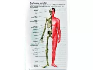
Arthritis
Arthritis. An algorithm. Radiographic evaluation of arthritis: inflammatory conditions Jon A. Jacobson, Gandikota Girish , Yebin Jiang, and Donald Resnick Radiology 2008 248:2, 378-389. Something different. Hypertrophic/proliferative Degenerative: primary or secondary

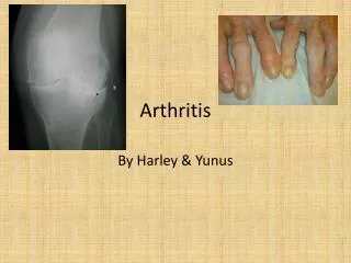
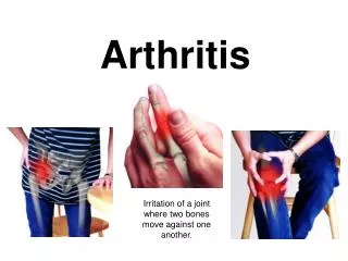
Arthritis
E N D
Presentation Transcript
An algorithm Radiographic evaluation of arthritis: inflammatory conditions Jon A. Jacobson, GandikotaGirish, Yebin Jiang, and Donald Resnick Radiology 2008 248:2, 378-389
Something different • Hypertrophic/proliferative • Degenerative: primary or secondary • Secondary AKA atypical OA • Hemophilia • Gout • trauma • Erosive • Rheumatoid + variants • Infectious • TB • Pyogenic/bacterial
Erosive • Synovial proliferation Pannus • Inflammatory erosions • Uniform joint space narrowing • Soft-tissue swelling • RA + seronegative variants • Reiters • Ankylosingspondylitis • Enteric arthropathy • Psoriatic arthropathy
RA • Multiple joints: systemic • Periarticularosteopenia • Juxtaarticular bony erosions (non-cartilage non-protected bone) • Subluxation and gross deformity • Periarticular soft tissue swelling
Cervical spine imaging in Ra Younes, Mohamed, et al. "Compared imaging of the rheumatoid cervical spine: prevalence study and associated factors." Joint Bone Spine 76.4 (2009): 361-368. The prevalence of rheumatoid cervical spine involvement was 47.5% by standard radiography, 28.2% by CT, and 70% by MRI. • Rheum involvement with high Sharp score and elevated CRP • 17.5% of those with Cspineabn were w/o sx. • Overall rheumatoid involvement of the cervical spine was not significantly associated with any of the epidemiological, clinical, laboratory, imaging, or therapeutic factors evaluated in the study.
DISH Criteria: SI and facet joints normal 4 contiguous vertebrae No disc space narrowing
hyperproliferative • Subchondral sclerosis, osteophytes • No erosions • Asymmetric joint space • Nearly allarthritides can lead to DJD *Weight-bearing radiograph for early detection Mobius battle
Microtrauma – not just body habitus but body habits AKA repeated use
GOUT Monu, Johnny UV, and Thomas L. Pope Jr. "Gout: a clinical and radiologic review." Radiologic Clinics of North America 42.1 (2004): 169-184.