ACL Risk Assessment Device for Athletes
130 likes | 230 Vues
Innovative diagnostic equipment for preventing ACL injuries in athletes through personalized training regimens based on biomechanical data. The device aims to enhance performance and reduce medical costs.

ACL Risk Assessment Device for Athletes
E N D
Presentation Transcript
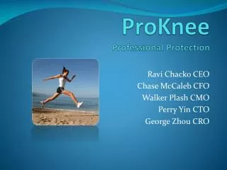
ProKneeProfessional Protection Ravi Chacko CEO Chase McCaleb CFO Walker Plash CMO Perry Yin CTO George Zhou CRO
Design Problem Each year in the US 100,000-200,000 people sustain a ruptured or torn ACL. (Mayo Clinic) Surgery >$10,000 Rehab >$10,000 Emotional, social and productivity costs
Known Risk Factors • Screening for risk factors has shown reduction in injury rates (Hewett) • Physiological Factors • Hormones, Sex, Leg Dominance, Joint Laxity/Recurvation • Behavioral Factors • Landing/Jumping • Limb position and resulting stresses on knee, hip, and ankle • Toe to heel shock absorption • Side-step/Cut • muscle firing patterns
Design Brief • Standardize ACL screening and preventative training • Method: • Measure leg mechanics (e.g. position, angle, forces) during jumping, landing or cutting • Determine risk factors and generate personalized training regimen to correct for those factors
Constraints and Specifications • Health and Safety • Should not overexert or injure user • Sensor device must be reusable • Cost • Customers will pay <$500 for this type of diagnostic equipment • Insurance may supplement cost • Reliability and Use • Results promptly displayed after testing • Imaging modality has sufficient resolution to determine position accurately • Calculations and results must yield significant medical benefit • Should allow freedom of movement for accurate results • Comfort • Device must be comfortable to wear with dimensions similar to athletic clothing • Cannot impede usual athletic movement
Existing Products Flock of BirdsMotion Capture Biodex System KINE Live Spring Loaded Goniometer
Components • Testing Modality • Landing, Cutting, Jumping • Sensors • Accelerometer • Electromyogram (EMG) • Goniometer • Gyroscopes • Pressure Sensor • Displacement Sensor • Output • Risk factors • Targeted training regimen • Live biofeedback
ProLand • Risk Factors Tested: • Knee Angle • Ankle Pronation • Components: • Gyroscopes • Pressure Sensor • Display
ProCut • Risk Factors: • Delay in activation of Vastus Lateralis (VL), Semitendinosis (ST) during sidecut • Components • EMG on VL and ST • Accelerometer/gyroscope • Display VL-ST delay: Risk Group: Zebis et al. 2009
ProScore • Risk Factors: • Knee and hip landing angle • Ankle and foot arch pronation • Ligament Dominance • Knee Abduction moment • Knee stability (Anterior-Posterior) • Components • Goniometers • Pressure Sensors • Gyroscopes • Wireless Transmitter • Display Hip Angle: Knee Angle: Medial Knee motion: Abduction Moment: Stability: Dynamic Foot Pronation: Ankle Pronation:
Thank you • Dr. Paul Sadja • Associate Professor of Biomedical Engineering • Jim Gossett • Associate Athletics Director for Sports Medicine • Paige Plash • Physical Therapist, COO Encore Rehabilitation • Dr. Elizabeth Hillman • Assistant Professor of Biomedical Engineering • Keith Yeager • Senior Staff Associate, Laboratory Manager