Download
1 / 73
760 likes | 1.09k Vues
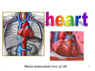
maria immaculata iwo, sf itb. BLOOD VESSEL. Blood vessels are of three types: arteries, capillaries, and Veins These vessels function to: 1. T ransport blood and its contents

E N D
maria immaculata iwo, sf itb BLOOD VESSEL
Blood vessels are of three types: • arteries, • capillaries, and • Veins These vessels function to: 1. Transport blood and its contents 2. Carry out exchange of gases in the pulmonary capillaries and exchange of gases plus nutrients for waste at the systemic capillaries 3. Regulate blood pressure; 4. direct blood flow to those systemic tissues that most require it at the moment.
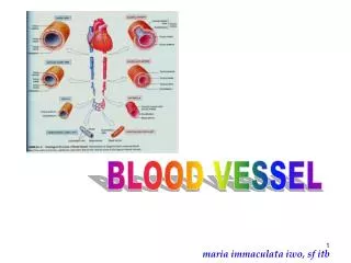
Darah didistribusi ke seluruh tubuh melalui pembuluh darah Arteri pembuluh eferent Membawa darah ke luar dari jantung Vena besar Vena cava: Superior - inferior JANTUNG aorta Arteri sedang Vena sedang Arteri kecil Arteri besar Vena kecil Arteriol Venula (vena terkecil) Capillary beds Vena pembuluh aferent Membawa darah kembali ke jantung Sistem limfatik Jaringan (Cairan interstisial)
Blood vessel circulates the blood to whole body Artery efferent vessel take blood away from heart Heart Big Vein Cava vein Superior & inferior Mdium Arteri Small Arteri Big Artery Medium Vein Arteriol Venules (smallest vein) Capillary beds Vein afferent vessel bring blood return to the heart Lymphatics System tissue (interstitial fluids)
Lapisan/dinding pembuluh darah • Tunika intima • Dinding dalam, • lapisan tipis tdd endothelium dan sedikit jar. ikat longgar, • sel-selnya berdekatan membentuk permukaan licin mengurangi friksi ketika darah melewati lumen pembuluh maria immaculata iwo, sf itb
Tunika media: • Lap. tengah, paling tebal, • tdd otot polos dan jar. elastik. • dikontrol oleh saraf simpatikus diameter pembuluh: • Konstriksi tek darah naik, • Dilatasi tek. darah turun • Tunika eksterna: • Lap. paling luar, tdd jar. ikat fibrous, • fungsi mensupport/melindungi pembuluh maria immaculata iwo, sf itb
ARTERY VEIN
ARTERIES • Arteries carry blood under great pressure, and their are adapted to handle that pressure Their relatively thick muscular walls make arteries elastic and contractile • Elasticity permits passive changes in vessel diameter in response to changes in blood pressure. • The contractility of the arterial walls enables them to change in diameter actively, primarily under the control of the sympathetic division of the autonomic nervous system. • When stimulated, arterial smooth muscles contract, thereby constricting the artery- a process called vasoconstriction. • Relaxation of these smooth muscle increases the diameter of the lumen – a process called vasodilation.
Wall of an artery consists of 3 distinct layers of tunics: Tunica intima • Composed of simple, squamous epithelium called endothelium. • Rests on a connective tissue membrane that is rich in elastic and collagenous fibers. Tunica media • Makes up the bulk of the arterial wall. • Includes smooth muscle fibers, which encircle the tube, and a thick layer of elastic connective tissue. Tunica adventitia • Is relatively thin. • Consists chiefly of connective tissue with irregularly arranged elastic and collagenous fibers. This layer attaches the artery to the surrounding tissues. • Also contains minute vessels (vasa vasorum vessels of vessels) that give rise to capillaries and provide blood to the more external cells of the artery wall.
Smooth muscles in the walls of arteries and arterioles are innervated by the sympathetic branches of the autonomic nervous system. Maria Immaculata Iwo, sf itb
arteries • Vasoconstiction and vasodilation affect : 1. The after-load on the heart 2. Peripheral blood pressure, and 3. Capillary blood flow. Contractility is also important during the vascular phase of hemostasis, when the contraction of damaged vessel wall helps reduce bleeding. maria immaculata iwo, sf itb
arteries • In traveling from the heart to peripheral capillaries, blood passes through • Elastic arteries Conducting arteries • Muscular Arteries Medium-sized arteries or distribution arteries • Arterioles (See Fig 21-2) maria immaculata iwo, sf itb
Elastic arteries • Elastic arteries, or conducting arteries, are large vessels with diameters up to 2.5 cm (1 in.) • These vessels transport large volumes of blood away from the heart. • The walls of elastic arteries (see Figure 21-2.) are extremely resilient because the tunica media contains a high density of elastic fibers and relatively few smooth muscle cells. • As a result, elastic arteries can tolerate the pressure changes that occur during the cardiac cycle. maria immaculata iwo, sf itb
Elastic arteries • During ventricular systole, pressures rise rapidly and the elastic arteries expand. • During ventricular diastole, blood pressure within the arterial system falls and the elastic fibers recoil to their original dimensions. • The elasticity of the arterial system dampens the pressure peaks and valleys that accompany the heartbeat. By the time, blood reaches the arterioles, the pressure oscillations have disappeared, and blood flow is continuous.
Muscular Arteries • Muscular arteries, also known as medium-sized arteries or distribution arteries, distribute blood to the body's skeletal muscles and internal organs. • Most of the vessels of the arterial system are muscular arteries. • These arteries are characterized by a thick tunica media that contains more smooth muscle cells than does the tunica media of elastic arteries (see Figures 21-1 and 21-2.). • A typical muscular artery has a lumen diameter of approximately 0.4 cm (0.16 in.), but some have diameters as small as 0.5 mm.
Muscular Arteries Superficial muscular arteries are important as pressure points - places in the body where muscular arteries can be forced against deeper bones to reduce blood flow and control severe bleeding. • The external carotid arteriesof the neck, • the brachial arteriesof the arms, • the mesenteric arteriesof the abdomen, • and the femoral arteriesof the thighs. are examples of muscular arteries Maria Immaculata Iwo, sf itb
Arterioles • Arterioles, with an internal diameter of 30 μm or less, smaller than muscular arteries. • Arterioles have a poorly defined tunica externa, • tunica media in the larger arterioles consists of one or two layers of smooth muscle cells The tunica media of the smallest arterioles contains scattered smooth muscle cells Maria Immaculata Iwo, sf itb
CAPILLARIES • Capillaries are the only blood vessels whose wall permit exchange between the blood and the surrounding interstitial fluids. • Because capillary walls are thin, diffusion distances are small, In addition, blood flows through capillaries relatively slowly, gave sufficient time for the diffusion or active transport of materials across the capillary walls. the histological structure of capillaries permits a two-way exchange of substance between blood and interstitial fluid. so exchange can occur quickly maria immaculata iwo, sf itb
CAPILLARIES • A typical capillary consists of an endothelial tube, a delicate basal lamina; neither a tunica media nor a tunica externa is present • The average diameter of a capillary is about 8 μm, very close to that of a red blood cell. • There are two major types of capillary: • Continuous capillaries • fenestrated capillaries.
Continuous Capillaries Are named because the cytoplasm of the endothelial cells is continuous when viewed in cross- section through a microscope. Cytoplasm appears as an uninterrupted ring, except for the endothelial junction. maria immaculata iwo, sf itb
Continuous Capillaries Most regions ofthe body are supplied by continuous capillaries. • Continuous capillaries are located in all tissues except epithelia and cartilage. • Continuous capillaries permit the diffusion of water, small solutes, and lipid-soluble materials into the surrounding interstitial fluid, but prevent the loss of blood cells and plasma proteins. • In addition, • some exchange may occur between blood and interstitial fluid by bulk transport the movement of vesicles that form through endocytosis at the inner endothelial surface. maria immaculata iwo, sf itb
Continuous Capillaries • In specialized continuous capillaries throughout most of the central nervous system and in the thymus, the endothelial cells are bound together by tight junctions. • These capillaries have very restricted permeability characteristics one example-the capillaries responsible for the blood-brain barrier maria immaculata iwo, sf itb
Fenestrated Capillaries Differ from continuous capillaries cells have numerous pores or fenestrations where the cytoplasm is very thin or absent. Found in kidneys, villi of the small intestine, chorroid plexus of the ventricles of the brain, and endocrine glands. Fenestrated capillaries (fenestra, window) are capillaries that contain "windows," or pores, that penetratethe endothelial lining (Figure 2l-4b).
Fenestrated Capillaries • The pores permit the rapid exchange of water and solutes as large as small peptides between plasma and interstitial fluid. • Examples of fenestrated capillaries include: the choroid plexus of the brain and the blood vessels in a variety of endocrine organs, such as the hypothalamus and the pituitary, pineal, and thyroid glands. Fenestrated capillaries are also located along absorptive areas of the intestinal tract and at filtration sites in the kidneys. Both the number of pores and their permeability characteristics may vary from one region of the capillary to another. maria immaculata iwo, sf itb
Sinusoids • Are wider than capillaries and • more tortuous • Contain spaces between • endothelial cells instead of having • the usual endothelial lining. • Basal lamina is incomplete or • missing. • In addition, sinusoids contain • specialized lining cells that are • adapted to the function of the • tissue. Sinusoids or Discontinuous Capillaries • In the liver, sinusoids contain phagocytic cells called stellate • reticuloendothelial (Kupffer) cells.
Sinusoids Sinusoids occur in the liver, bone marrow, spleen, and many endocrine organs, including the pituitary, parathyroid, and adrenal glands, and bone marrow. At liver sinusoids, plasma proteins secreted by liver cells enter the bloodstream. Along sinusoids of the liver, spleen, and bone marrow; phagocytic cells monitor the passing blood, engulfing damaged red blood cells, pathogens, and cellular debris. maria immaculata iwo, sf itb
Sinusoids Sinusoids resemble fenestrated capillaries that are flattened and irregularly shaped. In contrast to fenestrated capillaries, sinusoids commonly have gaps between adjacent endothelial cells, and the basal lamina thinner or absent. As a result, sinusoids permit the free exchange of water and solutes as large as plasma proteins between blood and interstitial fluid. • Blood moves through sinusoids relatively slowly; maximizing the time available for exchange across the sinusoidal walls. maria immaculata iwo, sf itb
Capillary Beds • Capillaries do not function as individual units but as part of an interconnected network called a capillary bed, or capillary plexus (Figure 21-5-). • A single arteriole generally gives rise to dozens of capillaries that empty into several venules the smallest vessels of the venous system. • The entrance to each capillary is guarded by a band of smooth muscle called a precapillarysphincter. • Contractionof the muscle cells narrows the diameter of the capillary, thereby reducing the flow of blood. • Relaxation of the sphincter dilates the opening, allowing blood to enter the capillary faster. maria immaculata iwo, sf itb
Figure 12.11 Anatomy of a capillary bed. • A capillary bed forms a maze of capillary vessels that lies between an arteriole and a venule. • When sphincter muscles are relaxed, the capillary bed is open, and blood flows through the capillaries. • When sphincter muscles are contracted, blood flows through a shunt that carries blood directly from an arteriole to a venule. • As blood passes through a capillary in the tissues, it gives up its oxygen (O2). Therefore, blood goes from being O2-rich in the arteriole (red color) to being O2-poor in the vein (blue color).
The cycling of contraction and relaxation of smooth muscles that changes blood flow through capillary beds is called vasomotion. • Vasomotion is controlled locally by changes in the concentrations of chemicalsanddissolved gases in the interstitial fluids. • For example, • when dissolved oxygen concentrations decline within a tissue, the capillary sphincters relax, so blood flow to the area increases. • This process, an example of capillary autoregulation, will be the focus of a later section. maria immaculata iwo, sf itb
When you are at rest, • blood flows through roughly 25% of the vessels within a typical capillary bed in your body. • Your cardiovascular system does not contain enough blood to maintain adequate blood flow to all the capillaries in all the capillary beds in your body at the same time. • As a results, • when many tissues become active, the blood flow through capillary beds must be coordinated. maria immaculata iwo, sf itb
Veins • Veins collect blood from all tissues and organs and return it to the heart. • The walls of veins can be thinner than those of corresponding arteries because the blood pressure in veins is lower than that in arteries. • Veins are classified on the basis of their size. Even though their walls are thinner, in general veins are larger in diameter than their corresponding arteries (Fig. 21-2, p.711) • Veins which carry blood back to the heart, follow pathways roughly parallel to those of the arteries. • Walls of veins are similar to those of arteries, in that they are composed of three distinct layers. • Middle layer is poorly developed. • As a result, veins have thinner walls that contain less smooth muscle and less elastic tissue than arteries.
Veins • Veins also function as blood reservoirs that can be drawn upon in time of need. • If a hemorrhage accompanied by drop in blood pressure occurs, the muscular walls of the veins are stimulated reflexively by the sympathetic nervous system. • Veins constrict and help to raise the blood pressure. • This mechanism ensures a nearly normal blood flow even if as much as 25% of the blood volume is lost. Maria Immaculata Iwo, sf itb
Venous Valves • Many veins, particularly • those in the arms and • legs, have flaps or valves • which project inward • from the lining. • Valves are usually • composed of two leaflets • that close if the blood • begins to back up in • the veins. • Valves are open as long as the blood flow is toward the heart • and closed if it is in the opposite direction.
Venous Valves • The arterial system is a high-pressure system. Almost all the force developed by the heart is required to push blood along the network of arteries and through miles of capillaries. • Blood pressure in a peripheral venule is only about 10 percent of that in the ascending aorta, and pressures continue to fall along the venous system. • The blood pressure in venules and medium-sized veins is so low that it cannot overcome the force of gravity. In the limbs, veins of this size contain valves, folds of the tunica intima that project from the vessel wall and point in the direction of blood flow. • These valves, like those in the heart, permit blood flow in one direction only. Venous valves prevent the back flow of blood. See FIG.21-6 The Function of Valves in the Venous System
Medium-Sized Veins Medium-sized veins range from 2 to 9 mm in internal diameter, comparable in size to muscular arteries. • In these veins, the tunica media is thin and contains relatively few smooth muscle cells. • The thickest layer of a medium-sized vein is the tunica externa, which contains longitudinal bundles of elastic and collagen fibers. Maria Immaculata Iwo, sf itb
Large Veins Large veins include the superior and inferior venae cavae and their tributaries within the abdomino pelvic and thoracic cavities. • All the tunica layers are present in all large veins. • The slender tunica media is surrounded by a thick tunica externa composed of a mixture of elastic and collagen fibers. Maria Immaculata Iwo, sf itb
Venules Venules are the microscopic vessels that continue from the capillaries and merge to form veins. Venules, which collect blood from capillary beds, are the smallest venous vessels. • They vary widely in size and structure. • An average venule has an internal diameter of roughly 20 μm. • Venules smaller than 50 μm lack a tunica media, and the smallest venules resemble expanded capillaries. maria immaculata iwo, sf itb
Blood Capillaries Water and other small molecules can cross through the cells of a capillary wall or through tiny clefts that occur between the cells. Large molecules in plasma, such as the plasma proteins, are too large to pass through capillary walls. • Three processes influence capillary exchange blood pressure, diffusion, and osmotic pressure:
Blood pressure, • which is created by the pumping of the heart, is the pressure of blood against a vessel’s (e.g., capillary) walls. • Diffusion, • as you know, is simply the movement of substances from the area of higher concentration to the area of lower concentration. Osmotic pressure - is a force caused by a difference in solute concentration on either side of a membrane.