Download
1 / 50
500 likes | 584 Vues
Water is vital for life, serving as a solvent, transporter, and participant in chemical reactions. It plays key roles in maintaining body composition and temperature regulation. Learn about its properties and functions in this comprehensive guide.

E N D
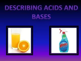
WATER, acids, bases, and buffers I. WATER Water is the solvent of life. It bathes our cells, dissolves and transports compounds in the blood, provides a medium for movement of molecules into and throughout cellular compartments, separates charged molecules, dissipates heat, and participates in chemical reactions. Most compounds in the body, including proteins, must interact with an aqueous medium function. In spite of the variation in the amount of water we ingest each day and produce from metabolism, our body maintains a nearly constant amount of water that is approximately 60% of our body weight.
A. Fluid Compartments in the Body Total body water is roughly 50 to 60% of body weight in adults and 75% of body weight in children. Approximately 40% of the total body water is intracellular and 60% extracellular. The extracellular water includes the fluid in plasma (blood after the cells have been removed) and interstitial water (the fluid in the tissue spaces, lying between cells). Transcellular water is a small, specialized portion of extracellular water that includes gastrointestinal secretions, urine, sweat, and fluid that has leaked through capillary walls because of such processes as increased hydrostatic pressure or inflammation.
B. Hydrogen Bonds in Water The dipolar nature of the water (H2O) molecule allows it to form hydrogen bonds, a property that is responsible for the role of water as a solvent. In H2O, the oxygen atom has two unshared electrons that form an electron dense cloud around it. This cloud lies above and below the plane formed by the water molecule.
In the covalent bond formed between the hydrogen and oxygen atoms, the shared electrons are attracted toward the oxygen atom, thus giving the oxygen atom a partial negative charge and the hydrogen atom a partial positive charge. As a result, the oxygen side of the molecule is much more electronegative than the hydrogen side, and the molecule is dipolar. Both the hydrogen and oxygen atoms of the water molecule form hydrogen bonds and participate in hydration shells. A hydrogen bond is a weak noncovalent interaction between the hydrogen of one molecule and the more electronegative atom of an acceptor molecule. The oxygen of water can form hydrogen bonds with two other water molecules, so that each water molecule is hydrogen-bonded to approximately four close neighboring water molecules in a fluid three-dimensional lattice.
WATER AS A SOLVENT Polar organic molecules and inorganic salts can readily dissolve in water because water also forms hydrogen bonds and electrostatic interactions with these molecules. Organic molecules containing a high proportion of electronegative atoms (generally oxygen or nitrogen) are soluble in water because these atoms participate in hydrogen bonding with water molecules. Chloride (Cl-), bicarbonate (HCO3-), and other anions are surrounded by a hydration shell of water molecules arranged with their hydrogen atoms closest to the anion. In a similar fashion, the oxygen atom of water molecules interacts with inorganic cations such as Na+and K+ to surround them with a hydration shell.
Although hydrogen bonds are strong enough to dissolve polar molecules in water and to separate charges, they are weak enough to allow movement of water and solutes. The strength of the hydrogen bond between two water molecules is only approximately 4 kcal, roughly l/20th of the strength of the covalent O-H bond in the water molecule. Thus, the extensive water lattice is dynamic and has many strained bonds that are continuously breaking and reforming. The average hydrogen bond between water molecules lasts only about 10 psec (1 picosecond is 10"12 sec), and each water molecule in the hydration shell of an ion stays only 2.4 nsec (1nanosecond = 10~9 sec). As a result, hydrogen bonds between water molecules and polar solutes continuously dissociate and reform, thereby permitting solutes to move through water and water to pass through channels in cellular membranes.
WATER AND THERMAL REGULATION The structure of water also allows it to resist temperature change. Its heat of fusion is high, so a large drop in temperature is needed to convert liquid water to the solid state of ice. The thermal conductivity of water is also high, thereby facilitating heat dissipation from high energy-using areas such as the brain into the blood and the total body water pool. Its heat capacity and heat of vaporization are remarkably high; as liquid water is converted to a gas and evaporates from the skin, we feel a cooling effect. Water responds to the input of heat by decreasing the extent of hydrogen bonding and to cooling by increasing the bonding between water molecules.
Osmolality and Water Movement Water distributes between the different fluid compartments according to the concentration of solutes, or osmolality, of each compartment. The osmolality of a fluid is proportionate to the total concentration of all dissolved molecules, including ions, organic metabolites, and proteins (usually expressed as milliosmoles (mOsm)/kg water). The semipermeable cellular membrane that separates the extracellular and intracellular compartments contains a number of ion channels through which water can freely move, but other molecules cannot.
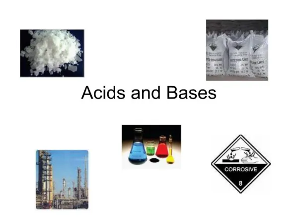
II. ACIDS AND BASES Acids are compounds that donate a hydrogen ion (H+) to a solution, and bases are compounds (such as the OH- ion) that accept hydrogen ions. Water itself dissociates to a slight extent, generating hydrogen ions (H+). Which are also called protons, and hydroxide ions (OH-). The hydrogen ions are extensively hydrated in water to form species such as H3O+, but nevertheless are usually represented as simply H+. Water itself is neutral, neither acidic nor basic. The pH of Water The extent of dissociation by water molecules into H+ and OH- is very slight, and the hydrogen ion concentration of pure water is only 0.0000001 M, or 10-7 mol/L. The concentration of hydrogen ions in a solution is usually denoted by the term pH, which is the negative logI0 of the hydrogen ion concentration expressed in mol/L. Therefore, the pH of pure water is 7.
The dissociation constant for water, Kd, expresses the relationship between the hydrogen ion concentration |H+|, the hydroxide ion concentration [OH-], and the concentration of water [H2O1 at equilibrium. Because water dissociates to such a small extent, [H2O] is essentially constant at 55.5 M. Multiplication of the Kd for water (approximately 1.8 X 10⁻¹⁶M) by 55.5 M gives a value of approximately 10 -l4 (M)2, which is called the ion product of water (Kw). Because Kw, the product of [H+] and [OH-], is always constant, a decrease of [H+] must be accompanied by a proportionate increase of [OH-].
A pH of 7 is termed neutral because [H+] and [OH-] are equal. Acidic solutions have a greater hydrogen ion concentration and a lower hydroxide ion concentration than pure water (pH<7.0), and basic solutions have a lower hydrogen ion concentration and a greater hydroxide ion concentration (pH>7.0). Strong and Weak Acids During metabolism, the body produces a number of acids that increase the hydrogen ion concentration of the blood or other body fluids and tend to lower the pH.
These metabolically important acids can be classified as weak acids or strong acids by their degree of dissociation into a hydrogen ion and a base (the anion component). Inorganic acids such as sulfuric acid (H2SO4) and hydrochloric acid (HC1) are strong acids that dissociate completely in solution.
Organic acids containing carboxylic acid groups (e.g., the ketone bodies acetoacetic acid and (β-hydroxybutyric acid) are weak acids that dissociate only to a limited extent in water. In general, a weak acid (HA), called the conjugate acid, dissociates into a hydrogen ion and an anionic component (A-), called the conjugate base. The name of an undissociated acid usually ends in "ic acid" (e.g., acetoacetic acid) and the name of the dissociated anionic component ends in "ate" (e.g., acetoacetate). The tendency of the acid (HA) to dissociate and donate a hydrogen ion to solution is denoted by its Ka, the equilibrium constant for dissociation of a weak acid. The higher the Ka, the greater is the tendency to dissociate a proton.
In the Henderson-Hasselbalch equation, the formula for the dissociation constant of a weak acid is converted to a convenient logarithmic equation. The term pKa represents the negative log of Ka. If the pK., for a weak acid is known, this equation can be used to calculate the ratio of the unprotonated to the protonated form at any pH. From this equation, you can see that a weak acid is 50% dissociated at a pH equal to its pKa. Most of the metabolic carboxylic acids have pKas between 2 and 5, depending on the other groups on the molecule. The pKa reflects the strength of an acid. Acids with a pKa of 2 are stronger acids than those with a pKa of 5 because, at any pH, a greater proportion is dissociated.
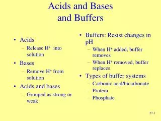
III. BUFFERS Buffers, which consist of a weak acid and its conjugate base, cause a solution to resist changes in pH when hydrogen ions or hydroxide ions are added. In figure, the pH of a solution of the weak acid acetic acid is graphed as a function of the amount of OH- that has been added. The OH- is expressed as equivalents of total acetic acid present in the dissociated and undissociated forms. At the midpoint of this curve, 0.5 equivalents of OH- have been added, and half of the conjugate acid has dissociated so that [A-] equals [HA]. This midpoint is expressed in the Henderson-Hasselbalch equation as the pKa, defined as the pH at which 50% dissociation occurs. As you add more OH- ions and move to the right on the curve, more of the conjugate acid molecules (HA) dissociate to generate H+ ions, which combine with the added OH- ions to form water. Consequently, only a small increase in pH results. If you add hydrogen ions to the buffer at its pKa (moving to the left of the midpoint), conjugate base molecules (A-) combine with the added hydrogen ions to form HA, and almost no decrease in pH occurs.
A buffer can only compensate for an influx or removal of hydrogen ions within approximately 1 pH unit of its pKa. As the pH of a buffered solution changes from the pKa to one pH unit below the pKa, the ratio of [A-] to HA changes from 1:1 to 1:10. If more hydrogen ions were added, the pH would fall rapidly because relatively little conjugate base remains. Likewise, at 1 pH unit above the pKa of a buffer, relatively little undissociated acid remains. More concentrated buffers are more effective simply because they contain a greater total number of buffer molecules per unit volume that can dissociate or recombine with hydrogen ions.
IV. METABOLIC ACIDS AND BUFFERS An average rate of metabolic activity produces roughly 22,000 mEq acid per day. If all of this acid were dissolved at one time in unbuffered body fluids, their pH would be less than 1. However, the pH of the blood is normally maintained between 7.36 and 7.44, and intracellular pH at approximately 7.1 (between 6.9 and 7.4). The widest range of extracellular pH over which the metabolic functions of the liver, the beating of the heart, and conduction of neural impulses can be maintained is 6.8 to 7.8. Thus, until the acid produced from metabolism can be excreted as CO2 in expired air and as ions in the urine, it needs to be buffered in the body fluids. The major buffer systems in the body are: the bicarbonate-carbonic acid buffer system, which operates principally in extracellular fluid; the hemoglobin buffer system in red blood cells; the phosphate buffer system in all types of cells; and the protein buffer system of cells and plasma.
Amino Acids in Proteins Proteins have many functions in the body. They serve as transporters of hydrophobic compounds in the blood, as cell adhesion molecules that attach cells to each other and to the extracellular matrix, as hormones that carry signals from one group of cells to another, as ion channels through lipid membranes, and as enzymes that increase the rate of biochemical reactions. The unique characteristics of a protein are dictated by its linear sequence of amino acids, termed its primary structure. The primary structure of a protein determines how it can fold and how it interacts with other molecules in the cell to perform its function. The primary structures of all of the diverse human proteins are synthesized from 20 amino acids arranged in a linear sequence determined by the genetic code.
I. GENERAL STRUCTURE OF THE AMINO ACIDS Twenty different amino acids are commonly found in proteins. They are all a-amino acids, amino acids in which the amino group is attached to the a-carbon (the carbon atom next to the carboxylate group). The α-carbon has two additional substituents, a hydrogen atom and an additional chemical group called a side chain (-R). The side chain is different for each amino acid.
At a physiologic pH of 7.4, the amino group on these amino acids carries a positive charge, and the carboxylic acid group is negatively charged. The pKa of the primary carboxylic acid groups for all of the amino acids is approximately 2 (1.8-2.4). At pH values much lower than the pKa (higher hydrogen ion concentrations), all of the carboxylic acid groups are protonated. At the pKa, 50% of the molecules are dissociated into carboxylate anions and protons, and at a pH of 7.4, more than 99% of the molecules are dissociated. The pKa for all of the a-amino groups is approximately 9.5 (8.8-11.0), so that at the lower pH of 7.4, most of the amino groups are fully protonated and carry a positive charge. The form of an amino acid that has both a positive and a negative charge is called a zwitterion. Because these charged chemical groups can form hydrogen bonds with water molecules, all of these amino acids are water-soluble at physiologic pH.
In all of the amino acids but glycine, the a-carbon is an asymmetric carbon atom that has four different substituents and can exist in either the D or L configuration. The amino acids in mammalian proteins are all L-amino acids represented with the amino group to the left if the carboxyl group is at the top of the structure. These same amino acids serve as precursors of nitrogen-containing compounds synthesized in the body, and thus human amino acid metabolism is also centered on L-amino acids. The amino acid glycine is neither D nor L because the a-carbon atom contains two hydrogen atoms.
The chemical properties of the amino acids give each protein its unique characteristics. Proteins are composed of one or more linear polypeptide chains containing hundreds of amino acids.
The names of the different amino acids have been given three-letter and one-letter abbreviations. The three-letter abbreviations use the first two letters in the name plus the third letter of the name or the letter of a characteristic sound, such as trp for tryptophan. The one-letter abbreviations use the first letter of the name of the most frequent amino acid in proteins (such as an "A" for alanine). If the first letter has already been assigned, the letter of a characteristic sound is used (such as an "R" for arginine). Single-letter abbreviations are usually used to denote the amino acids in a polypeptide sequence.
II. CLASSIFICATION OF AMINO ACID SIDE CHAINS The 20 amino acids used for protein synthesis are grouped into different classifications according to the polarity and structural features of the side chains.
GENERAL PROPERTIES The α-amino groups all have pK values near 9.4 and are therefore almost entirely in the ammonium ion form below pH 8.0. This leads to an important structural point: In the physiological pH range, both the carboxylic acid and the amino groups of a-amino acids are completely ionized. An amino acid can therefore act either as an acid or a base. Substances with this property are said to be amphoteric and are referred to as ampholytes (amphoteric electrolytes). Molecules that bear charged groups of opposite polarity are known as zwitterions or dipolar ions. The zwit-terionic character of the a-amino acids has been established by several methods including spectroscopic measurements and X-ray crystal structure determinations (in the solid state the a-amino acids are zwitter-ionic because the basic amine group abstracts a proton from the nearby acidic carboxylic acid group). Because amino acids are zwitterions, their physical properties are characteristic of ionic compounds.
Peptide Bonds The α-amino acids polymerize, at least conceptually, through the elimination of a water molecule. The resulting CO — NH linkage is known as a peptide bond. Polymers composed of two, three, a few (3-10), and many amino acid residues (alternatively called peptide units) are known, respectively, as dipeptides, tripeptides, oligopeptides, and polypeptides. These substances, however, are often referred to simply as "peptides." Proteins are molecules that consist of one or more polypeptide chains. These polypeptides range in length from ~ 40 to over 4000 amino acid residues.
Polypeptides are linear polymers; that is, each amino acid residue is linked to its neighbors in a head-to-tail fashion rather than forming branched chains.
CLASSIFICATION AND CHARACTERISTICS The most common and perhaps the most useful way of classifying the 20 "standard" amino acids is according to the polarities of their side chains (R groups). This is because proteins fold to their native conformations largely in response to the tendency to remove their hy-drophobic side chains from contact with water and to solvate their hydrophilic side chains. According to this classification scheme, there are three major types of amino acids: (1) those with nonpolar R groups, (2) those with uncharged polar R groups, and (3) those with charged polar R groups.
The Nonpolar Amino Acid Side Chains Have a Variety of Shapes and Sizes Nine amino acids are classified as having nonpolar side chains. Glycine (which, when it was found to be a component of gelatin in 1820, was the first amino acid to be identified in protein hydrolysates) has the smallest possible side chain, an H atom. Alanine, valine, leu-cine, and isoleucine have aliphatic hydrocarbon side chains ranging in size from a methyl group for alanine to isomeric butyl groups for leucine and isoleucine. Methi-onine has a thiol ether side chain, which resembles an n -butyl group in many of its physical properties (C and S have nearly equal electronegativities and S is about the size of a methylene group). Proline, a cyclic a-imino acid, has conformational constraints imposed by the cyclic nature of its pyrrolidine side group which is unique among the "standard " 20 amino acids. Phenylalanine, with its phenyl moiety, and tryptophan with its indole group, contain aromatic side groups, which are characterized by bulk as well as nonpolarity.
Uncharged Polar Side Chains Have Hydroxyl, Amide, or Thiol Groups Six amino acids are commonly classified as having uncharged polar side chains. Serine and threonine bear hydroxylic R groups of different sizes. Asparagine and glutamine have amide-bearing side chains of different sizes. Tyrosine has a phenolic group. Cysteine has a thiol group that is unique among the 20 amino acids in that it often forms a disulfide bond to another cysteine residue through the oxidation of their thiol groups.
Charged Polar Side Chains May Be Positively or Negatively Charged Five amino acids have charged side chains. The basic amino acids are positively charged at physiological pH values and comprise lysine, which has a butylammo-nium side chain, arginine, which bears a guanidino group, and histidine, which carries an imidazolium moiety. The acidic amino acids, aspartic acid and glu-tamic acid, are negatively charged above pH 3; in their ionized state, they are often referred to as aspartate and glutamate. Asparagine and glutamine are, respectively, the amides of aspartic acid and glutamic acid, as their corresponding carboxylates, aspartate and glutamate.
The allocation of the 20 amino acids among the three different groups is, of course, rather arbitrary. For example, glycine and alanine, the smallest of the amino acids, and tryptophan, with its heterocyclic ring, might just as well be classified as uncharged polar amino acids. Similarly, tyrosine and cysteine, with their ionizable side chains, might also be thought of as charged polar amino acids, particularly at higher pH values, while asparagine and glutamine are nearly as polar as their corresponding carboxylates, aspartate and glutamate.
Acid-Base Properties Amino acids and proteins have conspicuous acid-base properties. The a-amino acids have two or, for those with ionizable side groups, three acid-base groups. The titration curve of glydne, the simplest amino acid.
At low pH values, both acid - base groups of glycine are fully protonated so that it assumes the cationic form +H3NCH2COOH. In the course of the titration with a strong base, such as NaOH, glycine loses two protons in the stepwise fashion characteristic of a polyprotic acid. The pK values of glycine's two ionizable groups are sufficiently different so that the Henderson-Hasselbalch equation: closely approximates each leg of its titration curve. Consequently, the pK for each ionization step is that of the midpoint of its corresponding leg of the titration curve (Sections 2-2A and C): at pH 2.35 the concentrations of the canonic form, +H3NCH2COOH, and the zwitter-ionic form, +H3NCH2COO-, are equal and similarly, at pH 9.78 the concentrations of this zwitterionic form and the anionic form, H2NCH2COO-, are equal.
Amino acids never assume the neutral form in aqueous solution. The pH at which a molecule carries no net electric charge is known as its isoelectric point, pi. For the a-amino acids, the application of the Henderson -Hasselbalch equation indicates that, to a high degree of precision, where K, and Ky are the dissociation constants of the two ionizations involving the neutral species. For mono-amino, monocarboxylic acids such as glycine, Kiand Kj represent K1 and K2.
Proteins Have Complex Titration Curves The titration curves of the α-amino acids with ioniz-able side chains, such as that of glutamic acid, exhibit the expected three pX values. However, the titration curves of polypeptides and proteins, rarely provide any indication of individual pK values because of the large numbers of ionizable groups they represent (typically 25% of a protein's amino acid side chains are ionizable. Furthermore, the covalent and three-dimensional structure of a protein may cause the pK of each ionizable group to shift by as much as several pH units from its value in the free a-amino acid as a result of the electrostatic influence of nearby charged groups, medium effects arising from the proximity of groups of low dielectric constant, and the effects of hydrogen bonding associations. The titration curve of a protein is also a function of the salt concentration, as is shown in fig. because the salt ions act electrostatically to shield the side chain charges from one another, thereby attenuating these charge - charge interactions.
OPTICAL ACTIVITY The amino acids as isolated by the mild hydrolysis of proteins are, with the exception of glycine, all optically active; that is, they rotate the plane of plane-polarized light. Optically active molecules have an asymmetry such that they are not superimposable on their mirror image in the same way that a left hand is not superimposable on its mirror image, a right hand. This situation is characteristic of substances that contain tetrahedral carbon atoms that have four different substituents. The two molecules depicted are not superimposable since they are mirror images.
The central atoms in such atomic constellations are known as asymmetric centers, or chiral centers, and are said to have the property of chirality (Greek: cheir, hand). The Cα atoms of all the amino acids, with the exception of glycine, are asymmetric centers. Glycine, which has two H atoms substituent to its Cαatom, is superimposable on its mirror image and is therefore not optically active. Molecules that are nonsuperimposable mirror images are known as enantiomers of one another. Enantio-meric molecules are physically and chemically indistinguishable by most techniques.