Download
1 / 37
840 likes | 3.61k Vues
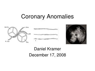
Coronary Artery Anomalies. Seoul National University Hospital Department of Thoracic & Cardiovascular Surgery. Coronary Artery Anomalies. Incidence : 1-2% of the population including minor (major anomalies<1% of CHD)

E N D
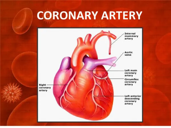
Coronary Artery Anomalies Seoul National University Hospital Department of Thoracic & Cardiovascular Surgery
Coronary Artery Anomalies • Incidence : 1-2% of the population including minor (major anomalies<1% of CHD) 1. Major anomalies 2. Minor variations - Coronary arterio-venous fistula - High take-off - Anomalous origin from PA - Anomalous circumflex artery origin - Single ostium or multiple ostia in Left coronary artery other aortic sinus Right coronaryartery - Hypoplastic proximal coronary artery Both coronary arteries - Congenital proximal or distal stenosis - Coronary artery from the posterior aortic sinus - Ventricular origin of an accessory coronary artery
Coronary Artery Anomalies • Major categories 1. Coronary arterio-venous fistula 2. Anomalous connection of left or right coronary artery to pulmonary artery 3. Anomalous origin of a main coronary artery from the aorta
Coronary Artery Anomalies • Pathophysiology • The anomalous origin of the left coronary artery on the main pulmonary artery leads to reversal of the left coronary arterial flow. This anomalous circuit results in coronary steal, myocardial ischemia, left ventricular dysfunction, and congestive heart failure. • Coronary arteriovenous fistulae between the left or right coronary arteries and the right heart chambers or the pulmonary artery also create a coronary steal phenomenon and myocardial ischemia.
Coronary Arterio-venous Fistula • Definition Direct communication between a coronary artery and the lumen of any one of the four cardiac chambers or the coronary sinus, or its tributary vein or SVC, pulmonary artery or pulmonary veins close to the heart • History Krause : 1st description in 1865 Trevor : 1st autopsy report in 1912 Biorck & Crafoord : 1st report of surgical correction in 1947 Fell : 1st report of correct diagnosis & surgical correction in 1958 Swan : 1st report of surgical correction by CPB in 1959
Coronary A-V Fistula • Morphology 1. Coronary artery site : dilated, elongated, serpiginous, Right coronary A : 50 ~ 55%, Left coronary A : 35% Both : 5% 2. Site of fistulous connection Right ventricle : 40% Right atrium : 25% Pulmonary artery : 15-20% Coronary sinus : 7% SVC : 1% Left atrium : 5% Left ventricle : 3% Other : rarely 3. Size & multiplicity of the fistula 2 ~ 5mm fibrous margin, single or multiple opening 4. Cardiac chamber : dilation in atrial, venous drain site 5. Bacterial endocarditis : 5% 6. Associated lesions : most occur as isolated lesions
Coronary A-V Fistula • Natural history • The fistula, if not present at birth, develops early in life. Likely small fistulae remains small and moderate fistulae slowly increase in size, although there may be little change over a 10 to 15 year period. • Onset of dyspnea, heart failure, and angina can occur in young patients with large fistula • The maximum incidence of congestive heart failure occurs in the fifth and sixth decades. • Spontaneous rupture and closure is very rare.
Coronary A-V Fistula • Clinical features & diagnosis 1. Presentation ;Most present late age in life 2. Symptoms ; Asymptomatic in young age Mild cardiomegaly Plethora on chest X-ray DOE from Lt. to Rt. shunt Angina (7%) Myocardial infarction (3%) Congestive heart failure (12 ~ 15%) in old age 3. Diagnosis ;Continuous murmurs EKG Chest radiography Echocardiography Cardiac catheterization, and angiography
Coronary A-V Fistula • Technique of operation 1.Closed without CPB When termination of a major coronary artery branch into an easily accessible site • When CPB is used Indications; (1) when artery is dilated & tortuous & relatively inaccessible as in AV groove (2) when the fistula is in the course of coronary artery (3) when an aneurysm requires excision Methods a. Closure through arteriotomy & aneurysm repair b. Closure through chamber
Coronary A-V Fistula • Surgical indications & results 1. Survival ◎ Early death Hospital mortality : rare Complication : rare ◎ Time related Late results : excellent 2. Indications Diagnosis is an indication for operation unless the shunt is very small(QP/QS<1.3) 3. Controversies Interventional catheter-delivered occluding devices and coils have been reported successfully
Anomalous Connection of Left Coronary Artery to Pulmonary Artery 1. Definition The whole of the left main or only the left anterior descending or circumflex branch connects anomalously to proximal main pulmonary artery, or very rarely to proximal right pulmonary artery. 2. History Brooks : 1st description in 1886 Bland, White & Garland (BWG Syndrome) : Described clinical syndrome in 1933 Sabiston : 1st successful operation in 1959 Cooley : Bypass graft in 1966 Neches : Coronary transposition in 1974 Takeuchi : Tunnel repair in 1979
ALCAPA • Morphology 1. Connection ◎ Left or posterior cusp of pulmonary trunk, rarely right cusp ◎ Uncommonly circumflex branch connection anomalously ◎ Rarely only the LAD connects anomalously 2. Left ventricle ◎ Hypertrophied, dilated ◎ Diffuse fibrosis in subendocardial layer ◎ Secondary subendocardial fibroelastosis 3. Mitral valve incompetence ◎ Extensive fibrosis, calcification in the papillary muscle (dysfunction) ◎ Endocardial fibroelastosis in the mitral apparatus & LV (fusion & shortening of chordae tendinae)
ALCAPA (1) Anomalous Connection of Left Coronary Artery to Pulmonary Artery
ALCAPA • Clinical features & diagnosis 1. Infant presentation Symptoms may be recognized within a week or so of birth (high postnatal pulmonary artery pressure limits runoff into the pulmonary artery) EKG : Anterolateral infarct with Q waves & ST segment elevation, LVH Chest radiology : Cardiomegaly with interstitial pulmonary edema Echocardiogram Cineangiography 2. Adult presentation Collateral circulation is adequate to prevent infarction. When severe symptoms do not occur in infancy, presentation is often delayed to beyond 20 years of age. Some patients remain asymptomatic.
ALCAPA • Natural history 1. Incidence rare (0.26%) , 1 per 400 CHD 2. Survival 65% : die due to LV failure during the 1st year of life lf death does not occur during the 1st year, the hazard lessens considerably by Rich interarterial collateral Restrictive opening of left coronary artery Marked right coronary dominance Most patients who survive infancy continue to be a risk of death from chronic heart failure secondary to ischemic cardiomyopathy
ALCAPA • Techniques of operation • Construction of a two-coronary system Tunnel operation (Takeuchi Repair) Left coronary artery transfer Subclavian-left coronary artery anastomosis Other techniques for assisting reimplantation Extension by autologous flaps of aorta or pulmonary artery Mobilization with pulmonary button Coronary artery bypass grafting • Ligation of left coronary artery
ALCAPA • Goals of surgery • Establishing the 2-coronary system with direct aortic implantation • Situations where direct aortic implantation is not suitable 1. Anomalous orifice is located in the nonfacing sinus or in the lateral position on the left pulmonary sinus 2. The patient is older with well-developed collateral around the pulmonary sinus
ALCAPA • Tunnel Operation
ALCAPA • Implantation of Anomalous Artery
ALCAPA • Subclavian-left Coronary Artery Anastomosis
ALCAPA • Surgical results 1. Survival Early death : variable Time-related : good 2. Mode of death : acute cardiac failure 3. Incremental risk factors for premature death Status of left ventricular myocardium lmportant mitral incompetence 4. Functional status : generally good late postoperatively 5. Left ventricular function 6. Mitral incompetence 7. Effect of the nature of the postoperative coronary system 8. Conduit patency after the two-vessel repair 9. Right ventricular outflow obstruction after a tunnel repair
ALCAPA • Indications for operation • The diagnosis in an infant is an indication for urgent operation and this condition in older patients as well. • The mitral valve should be left alone when operation is performed in young patients, but in older patients mitral repair or replacement may be required.
ALCAPA • Special Situations & Controversies 1. Anomalous connection of the right coronary artery, circumflex coronary artery, or left anterior descending coronary artery to pulmonary trunk . Even rarer . Much less lethal . Operation is probably advisable. 2. Total anomalous connection of the coronary arteries to the pulmonary trunk . Very rarely . Symptoms appear within a few days of birth and death follows within 2 weeks. . Urgent operation should be carried out.
Anomalous Coronary Artery Connection to Aorta • Definition A condition in which either the main coronary artery arises from the aorta in a site other than the left coronary sinus or the aorta just above it ; or in which right coronary artery arises in a site other than the right coronary sinus or the sino-tubular junction (apparently clinical events are related to this condition only when the anomalously arising artery passes between aorta & pulmonary trunk) • History Cheitlin : Description of death of anomalous LCA in 1974 Roberts : Description of anomalous RCA in 1984
Anomalous Coronary Artery Connection • Morphology 1. Anomalous connection of left main coronary artery . Two coronary ostia are side by side in the right sinus mostly. . Single enlarged ostium in the right sinus near to or overlying commissure between right and left less commonly . Coronary artery may pass posteriorly between aorta & PA trunk 2. Anomalous connection of right coronary artery . More common than anomalous connection of the left main coronary artery . The anomalously connecting right coronary artery passes forward between the two great arteries. . Commonly seperated ostium, but uncommonly single ostium within left sinus or over the commissure
Anomalous Coronary Artery Connection • Restriction of coronary flow • Acute angulation at the coronary takeoff • Presence of the ostial ridge, slitlike orifice, • Stretching and compression of the intramural segment by the aortic valve commissure • Most recently, variable lateral luminal compression of the intramural trunk that worsens during systole • RCAs arise from the left sinus or ectopically in the right sinus.
Anomalous Coronary Artery Connection • Medical (expectant) treatment • Coronary bypass grafting • Implantation into the correct sinus from outside the aorta • Translocation of the pulmonary artery • The use of primary angioplasty in patients with myocardial infarcts • The use of stents to prevent compression • Transaortic modification of the origin and proximal portion of the ectopic or anomalous RCA
Anomalous Coronary Artery Connection • Clinical features & diagnosis • The natural history is controversial. • No characteristic clinical or electrocardiographic feature • Diagnosed by coronary cineangiography A search is made for objective evidence of reversible ischemia (A number of cases have been reported in which serious sequelae, including sudden death)
Anomalous Coronary Artery Connection • Surgical Strategies Anomalous connection of a coronary artery (ACCAA) to an incorrect sinus of Valsalva is a relatively rare congenital defect • Coronary reimplantation, • Unroofing intramural segment, • Coronary artery bypass grafting. • Reinplantation of pulmonary trunk
Anomalous Coronary Artery Connection • Compromised coronary blood flow • Compression of the intramural segment of the coronary artery between the pulmonary artery and aorta • Narrowing of the slit-like orifice to anomalous artery with increased aortic pressure • An acute angle of the takeoff of the coronary artery • Stenosis in intramural segment of the coronary artery, especially at the level of the commissure.
Anomalous Coronary Artery Origin • Operative indications & techniques 1. Indication Indications remain arguable, but reversible ischemia is an indication for operation. 2. Technique . Coronary bypass graft operation internal mammary artery saphenous vein graft . Reconstruction of proximal portion of the coronary artery (lntramural course) . Division and reimplantation 3. Results Generally good
Anomalous Coronary Artery Connection Unroofing Procedure
Coronary Ostial Stenosis • Developmental etiology 1. Congenital 1) Membranous obstruction 2) Hypoplasia 3) Atresia 2. Inflammatory 1) Takayasu’s : 8.5% 2) Kawasaki : 9% 3) Middle aortic syndrome
Coronary Artery Aneurysm • Development & diagnosis • The first pathologic description of a coronary artery aneurysmwas by Morgagni in 1761, and the first clinical case of coronaryartery aneurysm was reported by Bourgon in 1812. • Coronaryartery aneurysm is an uncommon disease, the main causes of coronary artery aneurysm include congenital, atherosclerosis,Kawasaki disease, complication of percutaneous transluminalcoronary angioplasty, and endocarditis. • On rareoccasions, a coronary artery aneurysm grows large enough tobe called giant coronary artery aneurysm, for which a precisedefinition is still lacking
Coronary Artery Aneurysm • Clinical features • Coronary artery aneurysm is defined as coronary dilation thatexceeds the diameter of normal adjacent segments or the diameterof the patient's largest coronary vessel by 1.5 times. • Coronary artery aneurysm is an uncommon disease, found in only0.15% to 4.9% of patients who undergo coronary angiography. • The complications of coronary artery aneurysm include thrombosis,embolization, rupture, and vasospasm • Operative procedures include coronaryfistula closure, underwent coronary artery aneurysm resection,embolectomy, and plastic repair of the aortic wall with partialexcision of the free aortic wall.