Acute Retinal Necrosis
Acute Retinal Necrosis

Acute Retinal Necrosis
E N D
Presentation Transcript
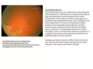
Acute Retinal Necrosis Acute Retinal Necrosis causes inflammation and subsequent retinal detachment. ARN presents in usually healthy patients with a periorbital pain, red eye (scleritis), then uveitis (inflammation of the anterior chamber) and progresses to peripheral retinal white/yellow spots, optic neuropathy, and retinal detachment. The cause is frequently the Herpes family of viruses especially Varicella (primary varicella, varicella zoster, and Ramsey-Hunt) infection and may be seen as part of the "uveomeningeal syndromes" (uveitis, meningitis). Since it is treated with intravenous acyclovir, it is a diagnosis that needs prompt attention and referral to an ophthalmologist or retinal specialist.Viewing acute retinal necrosis is difficult. Most of the time your view into the fundus will be obscured by the cellular response in the vitreous and anterior chamber. Acute Retinal Necrosis due to Herpes Zoster.The white arrow points to the optic disc.The arrow heads outline the area of necrosis in the retina.Photographs courtesy of Paul Zimmerman, MD
(B) ARN due to Herpes Zoster. The white areas are retinal necrosis.Photographs courtesy of Paul Zimmerman, MD (C) ARN due to Herpes Zoster.Here you see hemorrhages and white areas associated with ARN.Photographs courtesy of Paul Zimmerman, MD