Download
1 / 1
10 likes | 169 Vues
Tracking Severity and Distribution of Lung Disease During Mechanical Ventilation: Applications to Bronchoconstriction and Respiratory Distress Syndrome C. Bellardine 1 , E.P. Ingenito 2 , A. Hoffman 3 , F. Lopez 4 , W. Sanborn 4 , and K.R. Lutchen 1

E N D
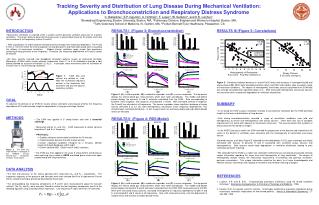
Tracking Severity and Distribution of Lung Disease During Mechanical Ventilation: Applications to Bronchoconstriction and Respiratory Distress Syndrome C. Bellardine1, E.P. Ingenito2, A. Hoffman3, F. Lopez4, W. Sanborn4, and K.R. Lutchen1 1Biomedical Engineering, Boston University, Boston, MA, 2Pulmonary Division, Brigham and Women's Hospital, Boston, MA, 3Tufts Veterinary School of Medicine, N. Grafton, MA, 4Puritan Bennett/Tyco Healthcare, Pleasanton, CA INTRODUCTION RESULTS I (Figure 3: Bronchoconstriction) RESULTS III (Figure 5: Correlations) • Mechanical ventilation is required when a patient cannot generate sufficient pressures to maintain ventilation. The lungs’ ability to generate these pressures is governed primarily by the elastic recoil and the resistance of the respiratory system (E and R). • With lung disease, R and E become elevated and increasingly more frequency dependent. The R and E from 0.1 to 8 Hz reflect the level and pattern of lung disease [1] and these data would aid in evaluating the efficacy of mechanical ventilation. Modern clinical ventilators apply simple flow waveforms containing energy primarily at one frequency. Therefore, the frequency dependence of R and E cannot be tracked. • We have recently invented new broadband ventilation patterns known as Enhanced Ventilator Waveforms (EVWs) which contain discrete frequencies (from 0.1 to 8 Hz) blended to provide a tidal breath followed by a passive exhalation [2] (Fig 1). In principle, these waveforms allow for estimation of R and E from 0.1 to 8 Hz during ventilation. Figure 1. EVW flow and volume are plotted vs. time. Note the enhanced frequency content in the inspiratory flow waveform and also the passive expiratory sections. • Figure 5. Correlation between decrease in arterial PaO2 levels and increases in heterogeneity (A) and airway closure (B). Within both bronchoconstriction (blue) and RDS (red) models, data revealed a range of constriction conditions. The degree of heterogeneity and airway closures quantified from EVW data was strongly correlated with significant drops in O2. Both increased heterogeneity and airway closures are consistent with a substantial degradation of ventilation distrubution. (A) (B) (C) Figure 3. (A) a mild responder, (B) a moderate responder, and (C) a severe responder. The top panels display the arterial blood gas measurements which were taken periodically. The middle and bottom panels display the dynamic R and E estimates calculated from the EVW. Data corresponding to baseline, initial response, final response, and albuterol is shown. With increased severity of response, the R and E are elevated at all frequencies. The severe responder shows significant evidence of airway closure (elevated E at 0.2 Hz) and heterogeneous constriction (more frequency dependence). This should impact ventilation distribution. In fact, oxygen levels are extremely depressed and carbon dioxide levels are elevated. GOAL SUMMARY To advance the delivery of an EVW for routine clinical ventilation and evaluate whether the frequency dependence of R and E provide insight on degradation of lung gas exchange function. • In all sheep the EVW sustains ventilation similarly to conventional ventilation but the EVW permitted insight on the level and distribution of lung disease. • Data during bronchoconstriction revealed a range of constriction conditions from mild and homogeneous to severe and heterogeneous with airway closures. Often there was lack of complete improvement in R and E with albuterol or recruitment maneuvers. This was consistent with a pattern of airway closures that would not reopen. • In the ARDS lung injury model, the EVW revealed the progression of the disease and showed that the extent of the defects in ventilation were consistent with the heterogeneity of constriction and airway closure. • Detailed analysis of all data (Figure 5) indicated that the degradation in PaO2 (gas exchange) was highly correlated with features of dynamic R and E associated with functional airway closures and heterogeneities. Both features would imply degradation in ventilation distribution leading to poor ventilation-perfusion matching. • We conclude that the EVW is a viable new ventilation method that can simultaneously provide clinically unique information regarding the mean level and heterogeneity of lung constriction. The degree of heterogeneity directly reflects the mechanical requirements of breathing and potential ventilation-perfusion mismatches. This unique information could be the basis to of more knowledgeable and effective clinician intervention with regards to treatment and ventilator weaning strategies. METHODS RESULTS II (Figure 4: RDS Model) • The EVW was applied in 5 sheep before and after a bronchial challenge. • Measured arterial O2 and CO2. EVW processed to obtain dynamic inspiratory R and E vs. frequency. • PROTOCOL: • Stabilize sheep on conventional ventilation for 15 minutes • Collect baseline dynamic R and E measurements • Deliver nebulized carbochol (16mg/ml for 2 minutes). Monitor response through blood gas and R and E. • Deliver albuterol MDI. Obtain final R and E estimates. • The EVW was then applied in the same 5 sheep before and during an oleic acid lung injury model of ARDS and blood gases were once again tracked along with lung mechanics. Figure 2. The EVW was implemented in a prototype of the NPB840 (Puritan Bennett/Tyco Healthcare) ventilator shown above. DATA ANALYSIS • The flow and pressure at the airway opening were measured (Qao and Pao, respectively). The inspiratory segments of the pressure and flow data were then isolated and fit to a trigonometric Fourier Series using the technique previously described by Kaczka [2]. • The corresponding low frequency components of R and E were recalculated to adjust for transient artifacts. The Qao and Pao were low pass filtered to isolate the low frequency components and fit to the following equation using a standard linear regression. Low frequency R and E were then re-estimated. REFERENCES (A) (B) (C) • Lutchen, K.R. and B. Suki. “Understanding pulmonary mechancis using the forced oscillation technique.” Bioengineering Approaches to Pulmonary Physiology and Medicine. 1996. • Kaczka, D.W., E. Ingenito, and K.R. Lutchen. “A technique to determine inspiratory impedance during mechanical ventilation: Implications for flow limited patients.” Annals of Biomedical Engineering. 27: 340-355, 1999. Figure 4. (A) a mild responder, (B) a moderate responder, and (C) a severe responder. The top panels display the arterial blood gas measurements which were taken periodically. The middle and bottom panels display the dynamic R and E estimates calculated from the EVW. With increased severity of RDS, there were increased airway closures, increased heterogeneity or frequency dependence of both R and E, and elevated R and E values at all frequencies. Also, with increased severity, note the depression of oxygen levels and increase of carbon dioxide levels.