The Foot
410 likes | 973 Vues
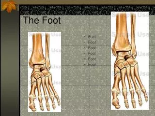
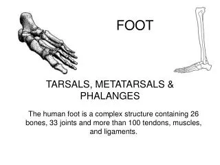
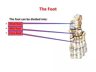
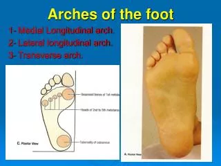
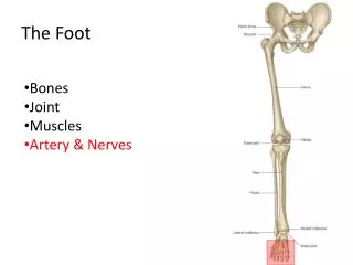
The Foot . Bones Joint Muscles Artery & Nerves. Superficial veins. Great s aphenous vein. Small saphenous vein. A rteries. Ant. Tibial. Dorsalis pedis artery. Ant. Tibial. Post. Tibial. Medial plantar Crosses over the two tendons long flexor Lateral plantar

The Foot
E N D
Presentation Transcript
The Foot • Bones • Joint • Muscles • Artery & Nerves
Ant. Tibial Dorsalispedis artery
Post. Tibial • Medial plantar • Crosses over the two tendons long flexor • Lateral plantar • Deep to flexor digitorum brevis
Common plantar digital n. Medial plantar n. • Flexor digitorum brevis • Flexor hallucis brevis • Abductor hallucis
Lateral plantar n. • Flexor accessories • Abductor digitiminimi • Interosseous muscles (deep brunch) • Adductor hallucis(deep brunch) • Flexor digitiminimibrevis(sup. brunch)
Surface Anatomy - Anterior, Extended Patella Indented Hollow
Surface Anatomy - Anterior, Flexed Patella Tibial Tuberosity Head Of Fibula
Palpation – Anterior* Patella: Lateral and Medial Patellar Facets Superior And Inferior Patellar Facets Medial Fat Pat Lateral Fat Pad Patellar Tendon**
Surface Anatomy - Medial Patella Tibial Tuberosity Medial Femoral Condyle Joint Line Medial Tibial Condyle
Palpation - Medial Medial Collateral Ligament (MCL)* Pes anserine bursa** Medial joint line
Surface Anatomy – Lateral Patella Quadriceps Tibial Tuberosity Head Of Fibula
Injury to the common peroneal nerve • The common fibular nerve may be severed during fracture of the fibula neck. • Results in paralysis of all muscles in the anterior and lateral compartments of the leg. • The loss of eversion of the foot and dorsiflexion of the ankle causes foot-drop. • Foot-drop: the foot drops and the toes drag of the floor when walking.
PNS Throughout Life • Dermatome – an area of skin • Innervated by cutaneous branches of a single spinal nerve • Embryonic muscles migrate to new locations • Some skin dermatomes become displaced • Muscles and skin always retain their original nerve supply
Posterior Anterior Map of Dermatomes
Innervation of the Skin: Dermatomes • Upper limb – skin is supplied by nerves of the brachial plexus • Lower limb: Lumbar nerves – anterior surface Sacral nerves – posterior surface
Elsie (L.3 ) is trying to rescue her clumsy man Slim (SI) from a septic tank(SciaTIC nerve), using a rope and a balloon. He has some GLUe (nerves to GLUteus muscles) on his leg. She is pregnant· is FEMale (FEMoral nerve) and has an OBstetric condition (OBturator nerve).
Lower limb dermatomes • L1 Dermatome:over trochanter and groin • L2 Dermatome:front of thigh to knee • L3 Dermatome:upper buttock, anterior thigh and knee, medial lower leg • L4 Dermatome:lateral Buttock, lateral thigh, medial leg, dorsum of foot, big toe • L5 Dermatome:Buttock, posterior and lateral thigh, lateral aspect of leg, dorsum of foot, medial half of sole, first, second, and third toes • S1 Dermatome: Buttock, thigh, and posterior leg • S2 Dermatome: Buttock, thigh, and posterior leg • S3 Dermatome: Groin, medial thigh to knee • S4 Dermatome: Perineum, genitals, lower sacrum