The Foot
The Foot. Foot Anatomy. The foot has many articulations which makes it a complex bone and soft tissue structure that undergoes a great deal of stress. Most athletic trainers will hear more complaints about this region than any other except the ankle. Osseous Structures.


The Foot
E N D
Presentation Transcript
Foot Anatomy • The foot has many articulations which makes it a complex bone and soft tissue structure that undergoes a great deal of stress. • Most athletic trainers will hear more complaints about this region than any other except the ankle.
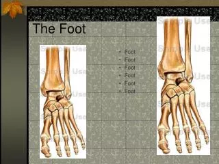
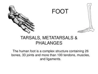
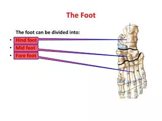
Osseous Structures • The foot is composed of 26 bones • It is subdivided into three regions • Hindfoot • Midfoot • Forefoot
Hindfoot • This region includes • The talus • The calcaneus • And their midtarsal articulations with the navicular and cuboid bones
Midfoot • This region includes • The navicular • The cuboid • And the 3 cuniform bones (numbered medial to lateral)
Forefoot • This region is comprised of • The metatarsals • And the phalanges • Not included in the 26 bones are the 2 sesamoid bones • These are the floating bones at the base of the great toe.
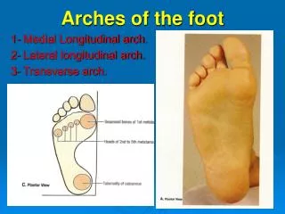
Arches • There are 2 arches in the foot • Transverse arch • Longitudinal arch • These are both held together by ligaments (static) and musculotendon units (dynamic).
Medial Longitudinal Arch • Consists of the calcaneal tuberosity, the talus, the navicular, three cuneiforms, and the 1st , 2nd, and 3rd metatarsal bones • It is maintained by the tibialis anterior, tibialis posterior, flexor digitorum longus, flexor hallucis longus, abductor hallucis, and the flexor digitorum brevis muscles • The ligaments included are the long plantar fascia and the plantar calcaneonavicular ligament.
Tibalis Posterior Tibialis Anterior
Lateral longitudinal arch • Is make up of the calcaneus, cuboid, and the 4th and 5th metatarsal bones • This arch is more stable and less adjustable than the MLA. • It is maintained by the peroneus longus, peroneus brevis, peroneus tertius, abductor digiti minimi, and flexor digitorum brevis muscles • The ligaments included are the long plantar ligament and the short plantar ligament
Transverse Arch • Is maintained by the tibialis posterior, tibialis anterior, and the peroneus longus muscles, and the plantar fascia • It consists of the navicular, cuneiforms, cuboid, and metatarsal bones. • It is divided into 3 parts • Tarsal • Posterior metatarsal • Anterior metatarsal • A loss in the anterior metatarsal arch results in callus formation under the heads of the metatarsal bones.
Articulations • Subtalar • Found in the hindfoot • It is a synovial joint having 3° of freedom • Resting position- Midway between the extremes of range of motion • Closed packed position- supination • Movements possible- gliding and rotation • Metatarsophalangeal • There are 5 of them; they are all synovial joints with 2 degrees of freedom. • Resting position- midway between extreme ranges of motion (10° extention) • Closed packed position- full extension • Movements possible- flexion, extention, abduction, and adduction
Interphalangeal joints • Synovial hinge joints with 1°of freedom • Resting position- Slight flexion • Closed packed position- Full Extension
Ligaments • Long Plantar • Lateral Retinaculum
Nerves and other structures • Peroneal nerve • Tibial nerve • Pedal pulse
Special Tests • Compression test (pott’s fracture)
Tinel’s Sign • Found in 2 places around the ankle • Anterior branch of the deep peroneal nerve • Posterior tibial nerve as it passes behind the medial malleolus • In both cases, tingling or paresthesia felt distally is a positive sign