Download
1 / 33
330 likes | 352 Vues
This module provides revision and update notes on liver disease, including liver size, consequences, sampling, necrosis, inflammation, viral infections, bacterial and parasitic infections, chronic liver disease, and neoplasia.

E N D
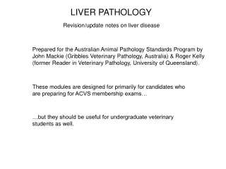
LIVER PATHOLOGY Revision/update notes on liver disease Prepared for the Australian Animal Pathology Standards Program by John Mackie (Gribbles Veterinary Pathology, Australia) & Roger Kelly (former Reader in Veterinary Pathology, University of Queensland). These modules are designed for primarily for candidates who are preparing for ACVS membership exams… …but they should be useful for undergraduate veterinary students as well.
LIVER PATHOLOGY Revision/update notes on liver disease Images from various sources have been used in these presentations, and every effort has been made to acknowledge these when they have come from sources other than our own files. We apologise for any inadvertent omissions or errors in attribution, and would be pleased to make appropriate corrections should these errors be brought to our attention.
Module 2 LIVER PATHOLOGY The second of 4 modules: • Module 1 • Background, normal structure, developmental anomalies • Functions of the liver • Failure of function (including clinical chemistry) • Module 2 • Consequences of liver size • Sampling (including normal cytology) • General pathological reactions • Necrosis • Module 3 • Inflammation • Circulatory disturbances • Final common pathway of chronic liver disease, incl. fibrosis • Viral infections • Module 4 • Bacterial infections • Parasitic infections • Acute hepatotoxicities • Chronic hepatotoxicities • Neoplasia
Module 2 Feline liver, ruptured in a road kill. Death was due to cranial trauma, not liver damage. Livers swollen by fatty change or neoplastic infiltrates are even more fragile, and are prone to spontaneous rupture, as in this case of equine hyperlipidaemia The size of the liver: its significance in liver disease The liver is large relative to most other solid organs. It constitutes 3-4% of body weight in adult carnivores, about 2% in omnivores, and about 1% in herbivores. The proportion of parenchyma relative to stroma is also large. So liver tissue is not very resistant to mechanical shearing forces… …and so liver is commonly ruptured during blunt abdominal trauma. Most cases of liver rupture resolve spontaneously and are unsuspected. This is because of the liver’s unspecialised distribution of function (disrupt a large part of the organ and the remainder can successfully take over its function)… …and blood lost into the abdomen is mostly resorbed via diaphragmatic lymphatics.
Module 2 Well-nourished ovine liver (left); malnutrition (right). Photomicrographs taken at same magnification. Hepatocyte cytoplasm atrophy; liver biochemistry within normal range. The size of the liver: its significance in liver disease Liver size and nutritional reserve The size of the liver, coupled with the fact that not all of it is needed to maintain liver function, has implications for nutritional reserve. A significant proportion of each hepatocyte can be “recycled” in times of severe malnutrition, thus providing emergency nutriment while sustaining near-normal liver function.
Module 2 The size of the liver: its significance in liver disease Liver size and endothelial surface area The size of the liver, coupled with its all-pervading sinusoidal vascular bed, means that the organ contains a vast endothelial surface area. Should this endothelium be extensively damaged, it will be unable to maintain the thrombosis-inhibiting mechanisms (plasminogen activation, etc) that are part of normal endothelial function in all tissues. This large area of damaged endothelium, in direct contact with passing blood, will immediately tend to attract platelets and activate the thrombotic cascade. As in the vasculature in general, in order that the affected liver sinusoids not become immediately completely thrombosed, this thrombotic cascade will in turn activate active thrombolysis so that sinusoidal blood can remain fluid and continue to flow.
Module 2 Splashy epicardial ecchymoses in acute fatal Cestrum poisoning in a cow (consumption coagulopathy secondary to extensive acute hepatic necrosis) The size of the liver: its significance in liver disease Liver size and endothelial surface area Because of the size of the liver; extensive sinusoidal endothelial damage can have the same depleting effects on prothrombin and fibrinogen concentration and platelet numbers as does disseminated intravascular coagulation, which is essentially the same process applied to the vasculature of the whole body. This depletion of clotting factors can be severe enough to cause spontaneous bleeding, which is why severe liver disease so often culminates in purpuric haemorrhage. The depletion of soluble clotting factors in this manner is compounded by the fact that most of them are synthesised in the liver, which, if failing, can of course no longer do so.
Module 2 Longitudinal section of canine hepatic venule (central vein) showing spiral configuration of the circular smooth muscle. The size of the liver: its significance in liver disease Liver size and blood volume Because the liver is so big and has such a voluminous sinusoidal vascular bed, it follows that a relatively modest increase in sinusoidal diameter across the whole organ can markedly increase the volume of blood within it. This is most dramatically illustrated in anaphylactic shock in dogs. In canids, the liver responds to anaphylaxis by constricting the spiral throttle muscles of hepatic venules, which causes instant sequestration of a large volume of blood within the sinusoids. This in turn results in paroxysmal hypovolaemia and catastrophic hypotension. At laparotomy the liver is intensely engorged and has coagulable lymph weeping from its capsule.
Module 2 Severe sinusoidal dilatation in bovine Pimelea poisoning. It is most severe in the periportal sinusoids (in contrast to passive venous congestion). Field case of Pimelea poisoning near Maree. The liver is much enlarged due to spectacular sinusoidal expansion. Blood dribbles from a capsular incision: the organ is like a huge bloody sponge The size of the liver: its significance in liver disease Liver size and blood volume In chronic passive hepatic congestion caused by congestive heart failure in cows, an increase of up to 13% in plasma volume was recorded in one study. The histology of chronically congested livers (see later) certainly shows marked sinusoidal distension, so this is hardly surprising. But for a truly spectacular illustration of plasma volume expansion involving the liver, try Pimelea spp. poisoning of cattle. Affected animals can have their total blood volume doubled, and their livers look like huge purple sponges. A combination of pulmonary hypertension and increased work-load on the heart leads eventually to heart failure.
Module 2 SAMPLING FOR LAB EXAMINATION Some general points regarding histological evaluation Ideally sample at least 2 regions of the liver (left and right) as streamlining of portal blood flow may lead to asymmetric distribution of lesions. Surgical biopsy techniques include wedge biopsy (at laparotomy), laparascopic biopsies using a spoon-like instrument (5 mm diameter core) and Trucut needle biopsies (1-2 mm diameter core). Liver Trucut needle biopsies have the advantage of sampling deeper within the liver parenchyma than wedges or laparoscopic biopsies. Disadvantages of Trucut biopsies include the small size of the biopsies (and thus reduced opportunity to examine entire acini) and the increased risk of tissue fragmentation and compression.
Module 2 SAMPLING FOR LAB EXAMINATION Some general points regarding cytological examination Cytology is most useful if there is generalised or lobar liver enlargement or if the liver contains a nodule or mass. It may also be useful for staging in cases of haemopoietic neoplasia (even when the liver is of normal size). Cytology is generally not useful if the liver is smaller than normal. The usual technique is percutaneous fine needle aspirate biopsy using a 22 G needle and 5 mL syringe. If diffuse liver disease is suspected, aspirate at least 2 regions of the liver (left and right) as streamlining of portal blood flow may lead to asymmetric distribution of lesions. If sampling a discrete nodule or mass, ultrasound-guided aspiration is recommended. Cytologic examination is often done to determine whether exploratory surgery is warranted.
Module 2 The background usually contains varying amounts of blood contamination with low numbers of leucocytes, mainly neutrophils. Pigment LIVER CYTOLOGY - NORMAL Fine needle aspirates of normal liver reveal clumps and sheets of oval to polyhedral cells, as well as single cells. Some normal hepatocytes may contain cytoplasmic dark blue granular pigment consistent with ceroid/lipofuscin. This needs to be differentiated from other pigments such as bile, haemosiderin and copper. Nuclei are round and centrally placed.
Module 2 LIVER CYTOLOGY - NORMAL Neutrophil Note clumps/sheets of oval to polyhedral cells with a relatively uniform nuclear:cytoplasmic ratio. Red cell Cytoplasm is abundant, pale-staining and slightly granular. The hepatocytes in this smear are normal. They could have been collected from a normal liver or from an unaffected area of a diseased liver. On the other hand, if collected from a mass, they could represent a hyperplastic nodule, hepatocellular adenoma, or even a well differentiated hepatocellular carcinoma. Hepatocyte
Module 2 Lamb; enterotoxaemia The pale, slightly elevated, sharply demarcated areas on this liver are caused by tiny gas bubbles accumulating during putrefaction. Note the difference between the right & left lobes. This might be due to the presence of more putrefactive saprophytes in the right lobe due to portal streaming. POSTMORTEM CHANGES These need to be differentiated from antemortem necrosis. Autolysis = self-digestion of cells and tissues by enzymes normally present in those tissues. Strictly speaking, this occurs in all tissues that die regardless of whether cells die before or after the animal dies (i.e. somatic death). Postmortem changes include both autolysis and putrefaction, the latter being the process by which bacteria break down tissues. Thus postmortem autolysis refers to the autolysis of cells occurring after somatic death. It is accelerated by bacterial decomposition. Postmortem change in the liver occurs rapidly because of its proximity to intestinal bacteria, which are released into the portal circulation at or just before death. The liver is also slow to cool, particularly in large animals. Grossly, postmortem change is characterised by softening of the substance of the liver, irregular pale foci on the capsular surface and formation of gas bubbles. There is dark staining of the parenchyma where the liver is in contact with intestine and around the gallbladder.
Module 2 POSTMORTEM CHANGES These need to be differentiated from antemortem necrosis histologically, too Microscopically, postmortem autolysis first affects hepatocytes in the periacinar zone (since these receive the least oxygen). Nuclear changes include shrinkage, minor fragmentation and then fading. There may be dissociation of hepatocytes and widening of sinusoids. Postmortem autolysis may be distinguished from antemortem necrosis by the lack of any inflammatory cell reaction. In addition, bacteria (mainly Clostridia of intestinal origin) may accompany the autolytic cells.
Module 2 The periacinar parenchyma on the left has been damaged more during the agonal period. AGONAL CHANGES Periacinar degeneration and necrosis may be seen in animals that have died rather slowly. During the prolonged agonal period, periacinar hepatocytes suffer hypoxia as a result of the failing circulation. This necrosis is more extensive if the animal is anaemic or has pre-existing pulmonary or thoracic pathology. Inflammatory cell reaction may be minimal and so distinction from postmortem autolysis may be problematic (especially if the liver is not fixed promptly after death). Affected hepatocytes tend to be shrunken, rounded, condensed and dissociated, and there may be loss of hepatocytes leading to disruption of the normal radiating cords.
Module 2 Severe ice-crystal artifact (yes, this is liver, in case you were wondering) Hepatocyte lysis caused by excessive barbiturate euthanasia solution OTHER ARTIFACTS 1. When tissues are frozen and then thawed before being placed in fixative, the formation of intracellular and extracellular ice crystals during freezing disrupts cells and tissues respectively. The extracellular ice crystals cause cleft-like spaces in subsequent histologic sections, which disrupt the tissue architecture and complicate microscopic evaluation. 2. The use of a barbiturate euthanasia solution of too high a concentration causes dissolution of hepatocytes, which also complicates microscopic evaluation.
HEPATOCELLULAR ATROPHY Module 2 Atrophy of hepatocytes results in smaller cells with reduced cytoplasm. Hepatic acini become smaller than normal, and if the change is diffuse the liver may appear grossly smaller, darker and firmer than normal. Atrophy may occur due to: Atrophic hepatocytes a. A lack of growth factors reaching the liver because of nutritional or circulatory factors (e.g. starvation or portosystemic shunt: see also Consequences of liver size: nutritional reserve Normal b. Local pressure from adjacent abdominal viscera (e.g. distended rumen in cattle or colon in a horse). This usually only affects one or two lobes of the liver. c. Chronic biliary tract disease with cholestasis (e.g. fascioliasis). This preferentially affects the left lobe in ruminants.
Module 2 Pale-staining nucleus (higher proportion of heterochromatin) Karyomegaly and cytoplasmic swelling (so-called megalocytosis) in ovine phomopsin poisoning. This example of cytomegaly shows most of the features of chronic hepatocellular damage including hydropic and fatty change. REACTIONS OF THE HEPATOCYTE TO INJURY 1. Hepatocyte enlargement (so-called megalocytosis) Chronic sublethal injury triggers a number of fairly non-specific changes in the appearance of hepatocyte cytoplasm and nucleus. Cytoplasm tends to increase in volume, due to (a) increase in water and solutes (hydropic degeneration), or (b) to increase in triglyceride (fatty change), or (c) to hypertrophy of endoplasmic reticulum, or (d) to various combinations of the above. Nuclei tend to enlarge (karyomegaly). Heterochromatin increases relative to euchromatin, so the nucleus stains paler overall in histological sections. The nucleolus/i become/s even more prominent.
REACTIONS OF THE HEPATOCYTE TO INJURY 2. Hydropic degeneration An early effect of hepatocellular injury is an increase in cytoplasmic water content This may be free in the cytosol or be more or less confined to membrane-lined channels and vesicles Hydropic degeneration may be reversible, provided organelle and cell membranes are not too badly damaged. Hydropic degeneration may be a precursor to cell death, in which case the hepatocyte is likely to burst (so-called lytic necrosis).
Severe hepatomegaly due to glucocorticoid hepatopathy in a dog. In this obese animal, fatty liver was a tempting diagnosis, but this was a case of pure glycogen storage. REACTIONS OF THE HEPATOCYTE TO INJURY 2. Hydropic degeneration Another form of hydropic change is in fact a post-mortem artifact. It is the result of excess glycogen accumulation in hepatocyte cytoplasm. The most extreme examples are seen in canine liver as a result of exposure to excess glucocorticoid. Immediately after circulation stops, the glycogen is transformed to glucose and then to lactic acid by anaerobic glycolysis in the still-living cell. These solutes, being osmotically active, draw water into the cytoplasm during the post-mortem interval, thus filling the cytoplasm with pale, irregular empty-looking spaces.
Module 2 Cytology - Normal Glycogen accumulation Hepatocytes are enlarged with vacuolated cytoplasm which is wispy and lucent. Note: no round droplet profiles.
Module 2 In this rapidly-fixed biopsy, the heavy glycogen deposits have been best preserved just beneath the surface, where fixation has inactivated glycolytic enzymes before the glycogen could be metabolised. Liver biopsy, glucocorticoid hepatopathy Glucocorticoid hepatopathy. The distribution of the vacuolation may be random, multifocal, or any one or combination of periportal, midzonal and periacinar, or diffuse involving all zones Deeper in the sample, the glycogen was converted to glucose and lactate before fixation occurred. REACTIONS OF THE HEPATOCYTE TO INJURY 2. Hydropic degeneration Glycogen accumulation and glucocorticoid (“steroid”) hepatopathy in dogs Histologically the hepatocytes are vacuolated and often markedly swollen. A significant feature of the vacuoles is that they never have sharp, round profiles like the lipid droplets seen in fatty liver (see below). The vacuoles may be PAS-positive, especially towards the periphery of the piece of tissue (e.g. under the capsule) where fixation occurred earliest. The severe cell swelling may perhaps cause canalicular compression or decreased sinusoidal blood flow. However, even severely affected animals rarely show clinical signs of liver damage. Many dogs with this disorder lack overt exposure to glucocorticoids, which suggests a role for chronic stress. There is no consistent relation between the magnitude of serum ALP activity, L-ALP activity, and the histological severity, though the vacuolation may precede increased serum ALP.
Glycogen storage in liver from a normal non-fasted rat. This has been called “feathery degeneration”, but it isn’t a pathological change. Module 2 Glycogen. Normal perfusion-fixed chicken liver Image courtesy Majid Ghoddusi GLYCOGEN ACCUMULATION Species variation Glycogen storage seems to be most spectacular in canids, but minor glycogen deposits are normal in well-fed animals and birds of most species.
Module 2 NORMAL LIPID METABOLISM Chylomicrons and free fatty acids are transported from intestine and adipose tissue respectively to liver. The fatty acids can be… …oxidised for energy by hepatocyte mitochondria (the liver derives most of its energy this way), or … … esterified to form triglycerides, which are then complexed with apoproteins to form very low density lipoproteins (VLDL) for secretion into plasma, or … … esterified with cholesterol for synthesis of cholesterol esters and subsequent export from the liver, or … … esterified with phosphate and carbohydrate moieties for synthesis of phospholipid.
Module 2 NORMAL LIPID METABOLISM Adipose tissue Triglycerides Dietary lipid (chylomicrons) Lipolysis (hormone sensitive lipase) Fatty acids Hepatocyte Export Fatty acids Triglycerides VLDL Storage B-oxidation Acetyl CoA Energy Ketones Mitochondria Export Energy Oxidation in peripheral tissues
Module 2 HEPATIC LIPIDOSIS = FATTY CHANGE You’d be surprised at the volume of triglyceride that traverses the normal liver in a day. It may approach the volume of the liver itself. The analogy with rush-hour city traffic is tempting: it is easy to envisage either excess input at certain times, as well as blockage of exit routes, and relatively mild disturbances of the system can lead to spectacular accumulations. Excess accumulation of triglycerides in hepatocytes. General mechanisms include: Increased entry of fatty acids, in excess of the ability of hepatocytes to utilise them. Examples include excess dietary intake, and mobilisation of fatty acids from adipose tissue stores due to high demand (e.g. starvation or diabetes mellitus). Liver fatty acid oxidation is maximal and excess acetyl CoA is shunted to the production of ketones (depending to some extent on the species). Increased synthesis of fatty acids (e.g. dietary carbohydrate excess). Decreased oxidation of fatty acids in mitochondria (e.g. hypoxia, some toxins). Decreased synthesis of apoproteins and thus decreased synthesis/export of lipoproteins (e.g. some toxins, protein deficiency). Decreased synthesis of phospholipid (e.g. choline deficiency).
Module 2 HEPATIC LIPIDOSIS = FATTY CHANGE Bovine abattoir specimen; fatty liver. A cow culled mid-lactation for foot disease. It is “normal” for heavily-lactating dairy cattle to have at least this fatty a liver. (an alternative name is steatosis) What are the gross features? The liver is enlarged, pale yellow, greasy, friable, and may float in water or fixative.
Module 2 Cytology of fatty liver normal lipidosis Many hepatocytes contain cytoplasmic vacuoles, which are nearly always round and sharply delineated (contrast with slide 22).
Module 2 Histology of fatty liver What are the histological features? Hepatocytes are enlarged with vacuolated cytoplasm. Vacuoles are round and sharply delineated. The nucleus is often displaced peripherally. The vacuoles may be large and single (macrovesicular fatty change) or small and multiple (microvesicular fatty change). There is little or no significance to the size or number of the vacuoles. It is the percentage of lipid in cytosol that counts.
Module 2 HEPATIC LIPIDOSIS = FATTY CHANGE Some specific causes: High triglyceride content (fatty liver) can be a physiological feature of liver; for example, in many avian species just after hatching during absorption of yolk sac. “Physiological” fatty liver can occur in situations of high energy demand such as late pregnancy, heavy lactation or peak egg production. Fatty liver strays into pathology when food intake falls suddenly for any reason in such high-production females, particularly if they are fat. Thus, fatty liver is always severe in pregnancy toxaemia and ketosis of ruminants. Diabetes mellitus: deficiency of (or resistance to) insulin causes accelerated mobilisation of fatty acids from adipose tissue stores, as the body burns more fat and tries to run on the ketones that are produced because glucose can’t be utilised. Sudden starvation in any fat animal. Hypoxia can affect fatty acid metabolism and protein synthesis. e.g. anaemia, cardiac failure, respiratory failure. Toxins that interfere with protein synthesis, thereby causing a bottleneck in export of lipoprotein into plasma for export to other tissues.
Module 2 Tension lipidosis adjacent to a site of capsular adhesion, in an otherwise normal bovine liver at slaughter. The subtlety of the circulatory compromise here shows how easy it is to tip the hepatocyte toward lipid accumulation. Ovine “white liver disease” Image courtesy Dr J King, Cornell University Hyperlipidaemia; obese pony HEPATIC LIPIDOSIS = FATTY CHANGE Some other specific examples: Equine hyperlipaemia and feline hepatic lipidosis.The pathogenesis is obscure but appears to involve a sudden onset of negative energy balance, perhaps simply precipitated by anorexia. Risk factors include obesity and (in horses) pregnancy or lactation. Relative insulin resistance may also play a role. Ovine white liver disease. The pathogenesis is obscure but appears to involve deficiency of cobalt/vitamin B12 in the face of lush pastures (“starvation in the midst of plenty”). Tension lipidosis.Local chronic hypoxia at or adjacent to the sites of capsular adhesions.
Module 2 Hypertrophied hepatocellular SER (chronic exposure to primidone). TEM Hypertrophy and aggregation of hepatocyte smooth endoplasmic reticulum in bovine liver; probably due to sublethal toxicity. Such material as this is likely to degrade and be taken up in autophagolysosomes Such material as this is likely to degrade and be taken up in autophagolysosomes. (image courtesy W. Castleman) OTHER HEPATOCELLULAR DEGENERATIONS Distressed cytoplasmic organelles: smooth endoplasmic reticulum Xenobiotics (toxic and non-toxic exogenous compounds absorbed from the gut) usually undergo some transformation by the liver, in the smooth endoplasmic reticulum (SER). This is the first stage in the process of preparing the substance for excretion. High intake of xenobiotic can cause hypertrophy of the SER. Sometimes the product of transformation is more toxic than the parent compound (see hepatotoxicity in module 3). If the hepatocyte is not killed, the hypertrophied SER can nevertheless be damaged. It tends to clump together and may become sequestered in phagolysosomes.