Download
1 / 66
660 likes | 888 Vues
Gastric Cancer. Zhejiang University. 浙江大学医学院附属第一医院 胃肠外科 于吉人. Ji-Ren Yu Department of GI Surgery The First Affiliated Hospital College of Medicine, Zhejiang University. Epidemiology. Jemal A, Bray F, Center MM, et al. Global cancer statistics. CA Cancer J Clin 2011; 61:69. Epidemiology.

E N D
Gastric Cancer Zhejiang University 浙江大学医学院附属第一医院 胃肠外科 于吉人 Ji-Ren Yu Department of GI Surgery The First Affiliated Hospital College of Medicine, Zhejiang University
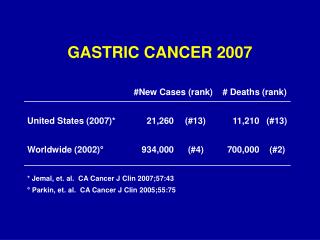
Epidemiology Jemal A, Bray F, Center MM, et al. Global cancer statistics. CA Cancer J Clin 2011; 61:69.
Epidemiology Jemal A, Bray F, Center MM, et al. Global cancer statistics. CA Cancer J Clin 2011; 61:69.
Risk Factors • 1. Nutrition • Low fat or protein consumption • Salted meat or fish • High nitrate consumption • High complex-carbohydrate consumption • 2. Environment and Heredity • Poor food preparation (smoked, salted) • Lack of refrigeration • Poor drinking water (well water) • Smoking
Risk Factors • 3.Social • Low socioeconomic status (except in Japan) • 4.Medical • Prior gastric surgeryHelicobacter pylori infection • Gastric atrophy and gastritisAdenomatous polyps • Pernicious anemia • Male gender
Etiological Factors (Risk Factors)
Correa mode of the pathogenesis of human gastric adenocarcinoma Pathology
Pathology • 1.Early gastric cancer (EGC) • Gastric cancer confined to the mucosa or submucosa, regardless of the presence or absence of lymph node metastasis 2. Advanced gastric cancer (AGC) Cancer cells infiltrate the proprial muscle layer or serosa
EGC Pathology IIb: superficially flat I: protruded IIc: superficially depressed IIa: superficially elevated III: excavated
EGC: Endoscopic images Type I Type II Type III
Pathology AGC: Borrmann’s classification Linitis plastica Borrmann'sclassification of gastric cancer based on gross appearance
T3 T4a T4b T1a T1b T4b T4a Lamina propria T1a T1b Subserosal connective tissue T stage are defined by depth of penetration into the gastric wall T stage
N stage Grouping of Regional Lymph Nodes (Groups 1-3) by Location of Primary Tumor According to the Japanese Classification of Gastric Carcinoma
Metastesis Direct invasion Lyphmatic metastesis Hematogenous metastasis Seeding metastasis
Clinical Presentation 1. Lacks specific symptoms early: vague epigastric discomfort indigestion. 2. Epigastric pain is constant, nonradiating, and unrelieved by food ingestion. 3. Advanced disease may present with weight loss, anorexia, fatigue, or vomiting. 4. Symptoms often reflect the site of origin of the tumor. Proximal tumors involving the gastroesophageal junction often present with dysphagia, whereas distal antral tumors may present as gastric outlet obstruction. 5. Hematemesis, anemic. 6. Very large tumors erode into the transverse colon, presenting as large bowel obstruction.
Physical signs 1. A palpable abdominal mass, 2. A palpable supraclavicular or periumbilical \lymph node, 3. Peritoneal metastasis palpable by rectal examination 4. A palpable ovarian mass (Krukenberg's tumor). 5. As the disease progresses, patients may develop hepatomegaly secondary to metastasis, jaundice, ascites, and cachexia.
Examination Endoscopy M-SCT (multiple detector-row spiral CT) BUS & EUS Double-contrast radiography MRI DL (diagnostic laparoscopy ) PET-CT
CT Clinicpathological Staging Laprascopy BUS EUS MRI PET-CT CTis the mainly procedure
Endoscopy Advanced carcinoma Carcinoma in situ
Niche Double-Contrast Barium Upper GI Radiography
EUS T N T
A B C N H1 T CT scan T4N2M1
BUS left right Liver metastasis Krukenberg’s tumor
Laparoscopy T T Abdominal metastasis
Treatment for Gastric Cancer Surgery Endoscopic mucosal resection (EMR) Endoscopic submucosal dissection (ESD) Laparoscopic Surgery Open Surgery Chemotherapy Chemoradiotherapy Target therapy
Criteria for EMR • NCCN 2011 V2. • 1.Early gastric cancer (Tis or T1a tumors limited) • 2. Well-differentiated or moderately differentiated histology • 3.Tumors less than 15mm in size, • 4.Absence of ulceration and no evidence of invasive finding • Japanese Gastric Cancer Association • Differentiated adenocarcinoma • Intramucosal cancer • 20 mm in size • without ulcer finding
Limitation of EMR techniques 1. Difficult to resect large than 20mm tumor in size 2. Difficult to resect ulcerative lesions ESD has been developed
ESD Oita Digestive Organs Hospital
ESD Oita Digestive Organs Hospital
Criteria for ESD NationalCancer Center Hospital In Japan
Principles of radical operation for gastric cancer 1. Negative margin (R0 resection, adequate margins ≥4 cm ) 2. D2 lymph node dissection for advance gastric cancer 3. Subtotal gastrectomy for distal gastric cancer 4.Total or proximal gastrectomy for proixmal gastric cancer Surgical Treatment for Gastric Cancer
Laparoscopic Resection 1. A suitable procedure for ECG (Our experience) 2. The efficacy and safety of this approach for advanc gastric carcinoma requires further investigation
Open Surgery for Advanced Gastric Cancer 1. A suitable procedure for ACG 2. R0 resection 3. R1 resection 4. R2 resection
Principles of advanced gastric cancer surgery Gastrectomy with regional lymphatics: perigastric lymph nodes(D1) and those along the named vessels of the celiac axis (D2), with a goal of examining 15 or greater lymph nodes Gastrectomy with D2 lymphadenectomyis the standard treatmentfor curable gastric cancer in eastern Asia
Gastrectomy and D2 lymphadenectomy for advanced gastric carcinoma Gastrectomy
Anastomosis Billroth II anastomosis Roux-en-Y anastomosis Subtotal gastrectomy
Left gastric A Hepatic A Splenic A No.11 LN