Download
1 / 101
1.08k likes | 1.6k Vues
Chromosomes, Mitosis and Meiosis. Chapter 10. Learning Objective 1. What is the significance of chromosomes in terms of information?. Chromosomes. Organization. Genes cell’s informational units made of DNA Chromatin DNA and protein makes up chromosomes (eukaryotes) Chromosomes

E N D
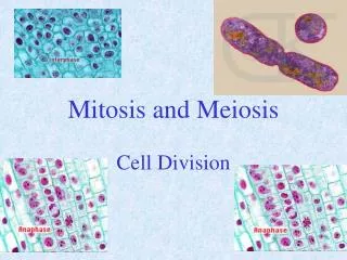
Chromosomes, Mitosis and Meiosis Chapter 10
Learning Objective 1 • What is the significance of chromosomes in terms of information?
Organization • Genes • cell’s informational units • made of DNA • Chromatin • DNA and protein • makes up chromosomes (eukaryotes) • Chromosomes • allow DNA sorting • into daughter cells
KEY CONCEPTS • In eukaryotic cells, DNA is wound around specific proteins to form chromatin, which in turn is folded and packaged to make individual chromosomes
Learning Objective2 • How is DNA organized in prokaryotic and eukaryotic cells?
Prokaryotic Cells • Contain circular DNA molecules
Eukaryotic Chromosomes • Nucleosome • histone (protein) bead wrapped in DNA • organized into coiled loops • held together by nonhistone scaffolding proteins
DNA wound around a cluster of histone molecules Histone tails Linker DNA Nucleosome (10 nm diameter) Fig. 10-2a, p. 213
100 nm Fig. 10-2b, p. 213
DNA Scaffolding proteins 2 μm Fig. 10-3, p. 213
1400 nm 300 nm fiber (looped domains) 700 nm 30 nm chromatin fiber DNA wound around a cluster of histone molecules Scaffolding protein Condensed chromosome Condensed chromatin Extended chromatin Packed nucleosomes Histone 10 nm Nucleosomes 2 nm DNA double helix Fig. 10-4, p. 214
Learning Objective3 • What are the stages in the eukaryotic cell cycle, and their principal events?
Eukaryotic Cell Cycle • Cycle of cell division • interphase • M phase
INTERPHASE G1 (First gap phase) S (Synthesis phase) G2 (Second gap phase) M PHASE (Mitosis and cytokinesis) Fig. 10-5, p. 215
Interphase • First gap phase (G1 phase) • cell grows and prepares for S phase • Synthesis phase (S phase) • DNA and chromosome protein synthesis • chromosome duplication • Second gap phase (G2 phase) • protein synthesis increases • preparation for cell division
M Phase • Mitosis • nuclear division • two nuclei identical to parent nucleus • Cytokinesis • cytoplasm divides • two daughter cells
KEY CONCEPTS • Cell division is an important part of the cell cycle, which consists of the successive stages through which a cell passes
Animation: The Cell Cycle CLICKTO PLAY
Learning Objective4 • What is the structure of a duplicated chromosome, including the sister chromatids, centromeres, and kinetochores?
A Duplicated Chromosome • Consists of a pair of sister chromatids • containing identical DNA sequences • Centromere • constricted region • joins sister chromatids • Kinetochore • protein to which microtubules bind • attached to centromere
Centromere region Microtubules Kinetochore Sister chromatids 1.0 μm Fig. 10-7, p. 218
Learning Objective5 • What is the process and significance of mitosis?
Mitosis • Preserves chromosome number • in eukaryotic cell division • Identical chromosomes are distributed to each pole of the cell • nuclear envelope forms around each set
INTERPHASE PROPHASE PROMETAPHASE Sister chromatids of duplicated chromosome Nucleolus Kinetochore Chromatin Nucleus Pieces of nuclear envelope Spindle microtubule Nuclear envelope Centrioles Developing mitotic spindle Plasma membrane Fig. 10-6a, p. 216
Prophase • Chromatin condenses into duplicated chromosomes (pair of sister chromatids) • Nuclear envelope begins to disappear • Mitotic spindle begins to form
Metaphase plate (cell’s midplane) Kinetochore microtubule (spindle microtubule) Centrioles Astral microtubules Pericentriolar material Polar (non- kinetochore) microtubule Sister chromatids Fig. 10-9a, p. 219
10 μm Fig. 10-9b, p. 219
Prometaphase • Spindle microtubules attach to kinetochores of chromosomes • Chromosomes begin to move toward cell’s midplane
Metaphase • Chromosomes align on cell’s midplane (metaphase plate) • Mitotic spindle is complete • Microtubules attach kinetochores of sister chromatids to opposite poles of cell
Anaphase • Sister chromatids separate • move to opposite poles • Each former chromatid is now a chromosome
Telophase • Nuclear envelope re-forms • Nucleoli appear • Chromosomes uncoil • Spindle disappears • Cytokinesis begins
METAPHASE ANAPHASE TELOPHASE 25 μm Spindle Cleavage furrow Centriole pair at spindle pole Reforming nuclear envelope Daughter chromosomes Cell’s midplane (metaphase plate) Fig. 10-6b, p. 217
KEY CONCEPTS • In cell division by mitosis, duplicated chromosomes separate (split apart) and are evenly distributed into two daughter nuclei
Cleavage furrow Actomyosin contractile ring 10 μm Fig. 10-10a, p. 220
Small vesicles fuse, forming larger vesicles Cell plate forming Nucleus Vesicles gather on cell’s midplane New cell walls (from vesicle contents) Eventually one large vesicle exists Plasma membrane Cell wall Cell plate forming New plasma membranes (from vesicle membranes) 5 μm Fig. 10-10b, p. 220
Learning Objective6 • How is the cell cycle controlled?
Cell-Cycle Control • Cyclin-dependent kinases (Cdks) • protein kinases that control cell cycle • active only when bound to cyclins • Cyclins • regulatory proteins • levels fluctuate during cell cycle