Download

1 / 104
1.08k likes | 1.57k Vues
Cranial Nerve Innervation of Ocular Structures. You’ve Got a Lot of Nerve(s)!. Introduction. Orbital structures are innervated by cranial nerves (CNs) II, III, IV, V, VI, and VII

E N D
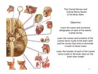
Cranial Nerve Innervation of Ocular Structures You’ve Got a Lot of Nerve(s)!
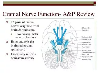
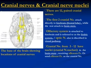
Introduction • Orbital structures are innervated by cranial nerves (CNs) II, III, IV, V, VI, and VII • Motor functions of the striated muscle are controlled by CN III, the oculomotor nerve; CN IV, the trochlear nerve; CN VI, the abducens nerve; and CN VII, the facial nerve • The trigeminal nerve, CN V, carries the sensory supply from the orbital structures • CN II, the optic nerve, carries visual information
The Nervous System • Afferent fibers bring information into the central nervous system (CNS)—these fibers generally have specialized nerve endings that respond to sensation producing stimuli such as touch, pressure, temperature, and pain • Information processing within the brain and spinal cord necessitates communication between different areas of the CNS through fiber tracts—a fiber tract may also be called a fasiculus, a peduncle, or a brachium • The portion of the cranial nerve from the cell body in the nucleus to its exit from the brain stem is the fasicular portion of the nerve
The Nervous System 2 • Efferent fibers, either somatic or autonomic, carry information from the CNS to target structures: muscles, organs, or glands • The efferent pathway in the somatic system usually consists of a fiber that runs the distance from the CNS to the target muscle—the autonomic pathway generally has a synapse within its pathway
Afferent Pathway: Orbital Sensory Innervation • The eye is abundantly supplied with sensory nerves that carry the sensation of touch, pressure, warmth, cold, and pain • Sensations from the cornea, iris, conjunctiva, and sclera consist primarily of pain—even light touching of the cornea registers as irritation or pain
Trigeminal Nerve • The fibers of the trigeminal nerve (CN V) serving ocular function are sensory and originate in the innervated structures • The description of the pathways of these nerves begins at a particular structure and follows the nerves as they join to become larger nerves, come together in the ganglion of the fifth cranial nerve, and then exit the ganglion and enter the pons • Considering the nerve in this manner, will mirror the direction of the action potential and information flow in these fibers • The trigeminal nerve consists of three branches: 1) ophthalmic, 2) maxillary, and 3) mandibular
Ophthalmic Division of CN V • The ophthalmic division of the trigeminal nerve, CN V, is formed from the following branches: • Nasociliary nerve • Frontal nerve • Lacrimal nerve • The maxillary division of the trigeminal nerve, CN V, is formed from the following branches: • Infraorbital nerve • Zygomatic nerve • The mandibular division of the trigeminal nerve innervates the lower face and contains both sensory and motor fibers—it enters the skull through the foramen ovale
Nasociliary Nerve • Sensory fibers from the structures in the medial canthus—caruncle, canaliculi, lacrimal sac, medial portion of the eyelids, the skin at the side of the nose—join to form the infratrochlear nerve • This nerve penetrates the orbital septum, enters the orbit below the trochlea, and runs along the upper border of the medial rectus • The anterior ethmoid nerve forms from fibers from the skin along the center of the nose, the nasal mucosa, and the ethmoid sinuses • The posterior ethmoid nerve forms from fibers from the ethmoid and sphenoid sinuses • Both nerves enter the orbit through the ethmoid foramina and join the nasociliary nerve along the orbit’s medial wall
Nasociliary Nerve 2 • Corneal innervation is dense (three to four time that of other epithelial tissue) and three networks of nerves are formed that are located in: • Corneal epithelium • Anterior stroma (subepithelial plexus) • Middle of the stroma • Nerves from these plexi come together in the peripheral stoma and radiate out into 70 to 80 branches, becoming myelinated in the last 2 mm of the cornea • Some of these branches join with nerves from other anterior segment structures to form two long ciliary nerves—one on the lateral side and one on the medial side of the globe
Nasociliary Nerve 3 • The long ciliary nerves run between the sclera and the choroid to the back of the eye and leave the eyeball at points approximately 3 mm on each side of the optic nerve • In addition to carrying afferent fibers out of the eye, they carry efferent sympathetic fibers into the eye to the dilator muscle • The long ciliary nerves then join up with the nasociliary nerve
Scleral Nerve Loops of Axenfeld • In some eyes (about 12%) the long ciliary nerve loops into the sclera from the suprachoroidal space, creating a dome-shaped elevation about 2 mm from the limbus on either the nasal or temporal side, but usually inferiorly • These are the scleral nerve loops (of Axenfeld) • Often this raised area is pigmented, usually blue or black, and should be differentiated from a melanoma • The nerve loop may produce pain when touched, which aids in its diagnosis
Nasociliary Nerve 4 • The other branches leaving the cornea join other sensory nerves and enter the choroid, join with the choroidal nerves, run to the back of the eye, where they leave as 6 to 10 short ciliary nerves • The short ciliary nerves exit the sclera in a ring around the optic nerve and enter the ciliary ganglion—the short ciliary nerves carry sympathetic and parasympathetic fibers in addition to sensory fibers • The sensory fibers do not synapse, but pass through the ganglion, leaving as the sensory root of the ciliary ganglion, which then joins the nasociliary nerve
Nasociliary Nerve 5 • In summary, the nasociliary nerve is formed by the joining of the infratrochlear nerve, the anterior and posterior ethmoid nerves, the long ciliary nerves, and the sensory root of the ciliary ganglion • The nasociliary nerve runs through the common tendinous ring and exits the orbit through the superior orbital fissure into the cranial cavity
Herpes Zoster • Herpes zoster (also knowns as shingles) is an acute CNS infection caused by the varicella-zoster virus and symptoms include pain and rash in the distribution area supplied by the affected sensory nerves • It seems the virus lies dormant in a sensory ganglion and, on becoming activated, migrates down the sensory pathway to the skin • An eruption of herpes zoster is more common in elderly people, but may occur at any age and may be related to a delayed hypersensitivity reaction
Herpes Zoster 2 • About 10% of all cases affect the ophthalmic division of the trigeminal nerve • Involvement of the tip of the nose often indicates that the eye will also be involved, reflecting the distribution of the nasociliary branches • This association of ocular involvement with zoster affecting the tip of the nose is called Hutchinson’s sign
Frontal Nerve • Sensory fibers from the skin and muscles of the forehead and upper eyelid come together and form the supratrochlear nerve, which enters the orbit by piercing the superior medial corner of the orbital septum • A second nerve forms in this same general area, the supraorbital nerve, lateral to the supratrochlear nerve • The supraorbital nerve enters the orbit as one or two branches: one branch enters through the supraorbital notch, along with the supraorbital artery • The supratrochlear and supraorbital nerves combine midway in the obit to form the frontal nerve, which runs back through the orbit between the levator and periorbita, and exits the orbit through the superior orbital fissure above the annulus of Zinn
Lacrimal Nerve • Sensory fibers from the lateral part of the upper eyelid and temporal region join and enter the lacrimal gland—they join the sensory fibers that serve the gland itself to form the lacrimal nerve • The lacrimal nerve leaves the lacrimal gland and runs posteriorly along the upper border of the lateral rectus muscle • The nerve receives a branch from the zygomatic nerve containing the autonomic innervation of the lacrimal gland • The lacrimal nerve exits the orbit through the superior orbital fissure above the muscle cone
Ophthalmic Nerve Formation • After leaving the orbit, the nasociliary nerve, the lacrimal nerve, and the frontal nerve join to form the ophthalmic division of the trigeminal nerve (CN V) • The ophthalmic nerve then enters the lateral wall of the cavernous sinus • While in the lateral wall, the nerve receives sensory fibers from the oculomotor, trochlear, and abducens nerves • Some of these fibers probably carry proprioceptive information from the extraocular muscles
Maxillary Division of CN V • The maxillary division of the trigeminal nerve forms from the infraorbital and zygomatic nerves, and other nerves from regions around the orbit
Infraorbital Nerve • The infraorbital nerve, formed by sensory fibers from the cheek, upper lip, and lower eyelid, enters the orbit through the infraorbital foramen • It runs posteriorly through the infraorbital canal and groove—while it is in the maxillary bone, branches join from the upper teeth and maxillary sinus • As the nerve leaves the infraorbital groove it exits the orbit through the inferior orbital fissure and joins other fibers in forming the maxillary nerve
Referred Pain • Referred pain is pain felt in an area remote from the actual site of involvement; however, the two areas are usually connected by a sensory nerve network • Frequently, the pathways of the trigeminal nerve are involved in referred pain—a common example is the momentary severe bilateral frontal headache sometimes experienced when eating ice cream • An abscessed tooth can cause pain described as ocular pain and should be suspected when no orbital cause for pain can be detected • These two events likely occur because the overload of sensation carried by the infraorbital nerve is interpreted by the brain as coming from another area also served by the trigeminal nerve
Zygomatic Nerve • Sensory fibers from the lateral aspect of the forehead enter the orbit through a foramen in the zygomatic bone as the zygomaticotemporal nerve • Fibers from the lateral aspect of the cheek and lower eyelid enter the orbit through a foramen in the zygomatic bone as the zygomaticofacial nerve • These two nerves join to become the zygomatic nerve, which runs along the lateral wall of the orbit, exiting the orbit through the inferior orbital fissure and joining with the maxillary nerve
Maxillary Nerve Formation • Formed by the joining of the infraorbital nerve, the zygomatic nerve, and nerves from the roof of the mouth, upper teeth, gums, and mucous membranes of the cheek, the maxillary nerve traverses the area between the maxilla and sphenoid bone • As it passes near the pterygopalatine fossa, it receives some autonomic fibers from the pterygopalatine ganglion—these fibers are destined for the lacrimal gland (zygomatic n. to lacrimal n.) • The maxillary nerve enters the skull through the foramen rotundum
Mandibular Division of CN V • The mandibular nerve innervates the lower face and contains both sensory and motor fibers • It enters the skull via the foramen ovale
Trigeminal Nerve Formation • As these three divisions—the ophthalmic, maxillary, and mandibular—enter the skull, they run posteriorly within the lateral wall of the cavernous sinus and enter the trigeminal ganglion (gasserian ganglion, semilunarganglion), where they synapse • The ganglion, flattened and semilunar in shape, is located lateral to the internal carotid artery and the posterior part of the cavernous sinus • The motor fibers of the mandibular division, which innervate the muscles of mastication, pass along the lower edge of the ganglion—only the sensory fibers synapse within the ganglion
Trigeminal Nerve Formation II • The fibers then leave the trigeminal ganglion and enter the lateral aspect of the pons as either the sensory root or the motor root of the trigeminal nerve • The sensory root carries information from the structures of the face and head, including all orbital structures • After entering the brain stem, these fibers form an ascending and descending tract, both terminating in the sensory nuclei of the trigeminal nerve
Trigeminal Nerve Formation III • The ascending tract terminates in the principal sensory nucleus in the pons, it registers the sensations of touch and pressure • The descending tract, which carries pain and temperature sensations, courses through the pons and medulla to the elongated nucleus of the spinal tract • The tract extends into the second cervical segment of the spinal cord • Information is relayed from the trigeminal nucleus to the thalamus
Oculocardiac Reflex • The oculocardiac reflex consists of bradycardia (slowed heartbeat), nausea, and faintness and can be elicited by pressure on the globe or stretch of the extraocular muscles (e.g., during surgery) • Fibers from the trigeminal spinal nucleus project into the reticular formation near the vagus nerve nuclei and can activate vagus synapses, precipitating this reflex • The motor aspect of the reflex can be blocked by a retrobulbar anesthesia or intravenous or intramuscular atropine
Efferent Pathway: Motor Nerves • The cranial nerves that supply striated muscles of the orbit and adnexa are the: • Oculomotor nerve • Trochlear nerve • Abducens nerve • Facial nerve
Oculomotor Nerve: Cranial Nerve III • The oculomotor nerve innervates the superior rectus, medial rectus, inferior rectus, inferior oblique, and the superior palpebral levator muscles • It also provides a route along which the autonomic fibers travel to innervate the iris sphincter muscle, the ciliary muscle, and the smooth muscles of the eyelid
Oculomotor Nucleus • The oculomotor nucleus is located in the midbrain, ventral to the cerebral aqueduct, at the level of the superior colliculus • It extends in a column from the posterior edge of the floor of the third ventricle to the trochlear nucleus • A defined area or subnucleus within the oculomotor nucleus controls each muscle—the proposed areas are based primarily on animal models • The nuclei for the MR, IR, IO, and SR are located in both the left and right oculomotor nucleus—the nucleus for the levator is single and is located centrally in the caudal area • Fibers to the IR, IO, and MR supply the ipsilateral eye—fibers innervating the SR decussate and supply the contralateral eye
Oculomotor Nucleus II • The decussating fibers pass through the opposite superior rectus nucleus—so damage to the right oculomotor nucleus might result in bilateral SR involvement • The centrally placed caudal nucleus provides innervation for both levator muscles • An autonomic nucleus, the accessory third nerve nucleus, or Edinger-Westphal nucleus, supplies parasympathetic innervation to the ciliary and iris sphincter muscles—it is located in the rostral, ventral part of the oculomotor nucleus
Oculomotor Nerve Pathway • Fibers from each of the individual nuclei join, forming the fascicular part of the nerve that passes through the red nucleus and the cerebral peduncle • These fibers emerge from the interpeduncular fossa on the anterior aspect of the midbrain as the oculomotornerve • The nerve passes between the superior cerebellar and posterior cerebral arteries as it runs forward, lateral to, and slightly inferior to the posterior communicating artery of the circle of Willis • The nerve pierces the roof of the cavernous sinus and runs within its lateral wall above the trochlear nerve • While in the cavernous sinus the oculomotor nerve sends small sensory branches to the ophthalmic nerve and receives sympathetic fibers from the plexus around the internal carotid
Oculomotor Nerve Pathway II • The oculomotor nerve exits the sinus and enters the orbit through the superior orbital fissure, having divided into superior and inferior divisions—both divisions are within the oculomotor foramen • The superior branch runs medially above the optic nerve and enters the superior rectus on its inferior surface—additional fibers either pierce the muscle or pass around it to innervate the levator • The inferior branch runs below the optic nerve and divides into three branches
Oculomotor Nerve Pathway III • The first branch enters the MR on its lateral surface • The second branch enters the IR on its upper surface • The third branch gives off parasympathetic fibers that form the parasympathetic root to the ciliary ganglion—then it runs along the lateral border of the IR, crossing it to enter the IO muscle near its midpoint • These parasympathetic fibers arise in the Edinger-Westphal nucleus and synapse in the ciliary ganglion
Trochlear Nerve: Cranial Nerve IV • The trochlear nerve innervates the SO muscle • Its fibers travel dorsally and decussate • CN IV is the only cranial nerve to cross—so the trochlear nucleus innervates the contralateral SO muscle • The trochlear nucleus is located in the midbrain anterior to the cerebral aqueduct and below the oculomotor nucleus, at the level of the inferior colliculus