Download
1 / 41
480 likes | 1.24k Vues
A REVIEW OF DIABETIC FOOT. By Hinnah Nasreen. Diabetic Foot. A wound/ulcer located on weight bearing areas on foot(usually plantar surface over the metatarsal heads or on the heel) in diabetic patients. IMPORATANCE Preventable so patient education required

E N D
A REVIEW OF DIABETIC FOOT By HinnahNasreen
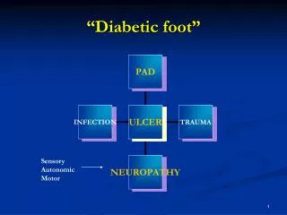
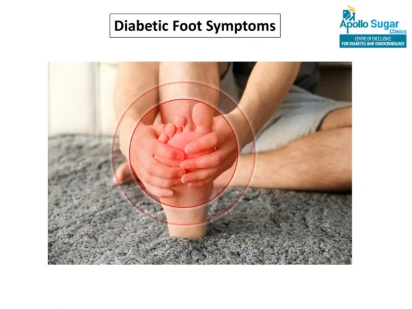
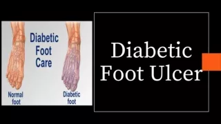
Diabetic Foot A wound/ulcer located on weight bearing areas on foot(usually plantar surface over the metatarsal heads or on the heel) in diabetic patients. IMPORATANCE • Preventable so patient education required • Consequences….. Major morbidity and deaths • Very common problem
Etiology Contributory Factors • Trophic changes from Peripheral neuropathy….. >80% • Ischemia due to atheroma of arteries… 60% • Low resistance to infection due to excess sugars in tissues • Poor vision • Limited mobility in joints • Cerebrovascular disease • PVD……> peripheral edema
I- NEUROPATHY Motor Sensory Autonomic ↓ Proprioception, Unawareness of foot position Muscle wasting Foot weakness Postural deviation A-V Shunt* open Permanent Reduced sweating Increase foot Blood flow Dry skin ↓ nociception Stress on bones & joints Plantar pressure Deformities, stress and shear pressures Fissures and cracks Bulging foot veins, Warm foot Callus formation Trauma Infection ULCER
II- Peripheral arterial disease Artheroma plaque narrowing the arterial lumen Artherosclerosis narrows or blocks the arterial lumen Foot ischaemia Necrosis /Gangrene Foot ulcer Ischemic toes due to atherosclerosis • PVD is difficult to detect as AV shunting appears and surrounding parts have good circulation until line of demarcation develops. Infection
III- Infection • Decreased resistance to infection due to • increased sugars in tissues and • Malfunction of lymphocytes. • Infection gains entry through callosities and amateur chiropody. • Infection involving fascia, tendon and bone can spread proximally via subfascialplane.
Most Common Isolates at Baseline(50% had only Gram positive cocci isolated) Lipsky BA, et al. Clin Infect Dis.2004;38.
Common Foot Ailments and Deformities 3. Onychogryphosis= Thickened (Ram’s horn) nail TOENAILS 2. Onychocryptosis= ingrown nail 1. Onychomycosis= Fungal nails
4. Onycholysis= crumbly nail 5. Onychia/ Paronychia= abscessed/ infected nail 6. Xerosis including cracked heals
corn calluses 7. Corns/Calluses 8. HalluxAbductovalgus AKA Bunions
9. Hammer Toes (hammer digit syndrome) 10. Claw Toes
11. Pescavus or High arch feet Normal High arch
12. Charcot Foot • This is where bone and joint destruction occurs.
History Diagnosis • Glycemic control • Duration of diabetes • Renal disease • Cigarette smoking • Poor social circumstances • Clinical Assessment of Foot • Skin & Integument • Vascular • Neurological • Musculoskeletal • Shoegear
SKIN AND INTEGUMENT • Color • Temperature • Texture • Hair Growth • Moisture • Lesions-location, size, type • Condition of toenails • condition of subcutaneous padding • Ability to pass a probe directly to underlying bone is a sensitive marker of osteomyelitis.
VASCULAR EXAMINATION • Color • CFT • Varicosities/Edema • Pulses (DP, PT, popliteal and femoral) • Ankle/ brachial pressure index • Presence of abdominal and femoral bruits
VASCULAR EXAMINATION (contd) • Doppler scan ( to measure anterior and posterior tibial pressure) • Angiography ( for large vessel disease) • Transcutaneous oxygen pressure measurement
NEUROLOGICAL EXAMINATION • Light touch • Sharp-dull discrimination • Vibratory (Testing with biosthesiometer or tuning fork) • Proprioception • Protective sensation • DTR
DISCRIMINATORY TOUCH • Tested by Semmes-Weinstein (10gauge) monofilament is applied to high risk areas of foot. • The monofilament is placed perpendicular to the skin’s surface. • Apply sufficient force to cause the filament to bend or buckle, using a smooth, not jabbing motion. • The total duration of the contact at each site should be approximately 1 to 2 seconds.
MUSCULOSKELETAL EXAMINATION • General appearance(gross deformities) • Muscle strength/function • Muscle tone • ROM
SHOE GEAR • Everyday shoe style • Dress shoe style • Exercise shoes • Shoe inserts/orthoses • Special shoe needs • Inspect socks or hose for blood or other discharge • Examine footwear for torn linings, foreign objects, breathable materials, abnormal wear patterns, and proper fit.
Investigations • LABORATORY • Urine and blood sugar • c/s of discharge • Blood culture • RFT • Lipid profile • ECG • Echocardiography • Start empirical t/m before results
RADIOLOGICAL • CXR • For osteomyelitis • Plain X-ray….. In acute cases abnormal after 10- 40days • Bone scan…. ..+ive within 24hrs • MRI… ………….access extent detailed anatomical info • Bone biopsy…. Chronic cases • Osteomyelitis is not an indication for amputation
Management • Preventive measures • Reduction or elimination of diabetic patients • Treatment of infection early • Correction of ischemia • Maintainence of environment • Promoting wound healing • Follow up • A multidisciplinary team approach is beneficial, including infectious disease, podiatry and/or orthopedic surgery, vascular surgery, endocrinology, and rehabilitation specialists.
Prevention PATIENT EDUCATION AND COMMUNITY CARE • Self management • Support of doctor • Counseling PATIENT’S CHECK LIST DAILY EXAMINATION • Look for cuts or sores • Check for warning signs….redness, swelling, warmth, pain, slow healing, dry cracks, bleeding corns or callus, loss of sensations
DAILY CARE • Wash daily • Dry carefully esp. web spaces • Use talcum powder • Do not cut corns or callus • Toe nails trimmed and smooth • Treat dry skin or athlete’s foot • Keep blood glucose under control • Periodic foot examination: once a year for every foot every 3-4 months for foot at risk Nail cutting
FOOT WEAR • Don’t walk bare foot • Don’t wear too tight shoes • Wear shoes with socks • Wear well cushioned shoes • Buy roomy shoes with fastening.. Lace or velcro • Don’t wear new shoes for more than half hour • For diagnosed cases special foot wear are available
Conservative Management REDUCTION OR ELIMINATION OF DIABETIC PATIENT • Control of diabetes by diet and drugs • Stop smoking • Reduce weight if over weight TREAT INFECTION • Antibiotics after c/s • Cover polymicrobial infection (gram –ive, +ive and anaerobes) SPECIFIC MANAGEMENT FOR ULCER • Debridement of infected tissues • Local wound care • Off loading
WOUND DEBRIDEMNT • Remove necrotic tissue that carries heaviest bacterial load • Keep tissues warm and moist to prevent formation of necrotic material • Alginates.. Topical dressing..20-30 times… more ability to absorb fluid • split-skin graft • Vacuum-Assisted closure (VAC) pump It applies gentle negative pressure to the ulcer via a tube and foam sponge which are applied to the ulcer over a dressing and sealed in place with a plastic film to create a vacuum. Exudate from the wound is sucked along the tube to a disposable collecting chamber. The negative pressure improves the vascularity and stimulates granulation of the wound.
WOUND DEBRIDEMNT (contd) • Dermagraft (cultured skin dermis) • Growth factor injected in edges of ulcer (if not healing) • Hyperbaric oxygen therapy • Larvatherapy Maggots are available in 2 forms. • ‘Free Range’ maggots • BioFOAMDressing
OFF LOADING PRESSURE: CASTS • Distributes weight bearing pressure over a large surface area • Optimal device is TCC • Total contact cast (TCC) • It is a close-fitting plaster of paris and fibreglass • minimum padding • efficient method of redistributing plantar pressure, and should be reserved for plantar ulcers that have not responded to other casting treatments. • Allow mobility while ulcer heals
Air cast (walking brace) • A bivalve cast with the halves joined together with Velcro strapping. • The cast is lined with 4 air cells which can be inflated with a hand pump to ensure a close fit. • The cast can be removed easily by patients to check their ulcers and before going to bed. • Scotch cast boot A simple, removable boot made of stockinette, soffban bandage, felt and fibreglass tape.
Removable cast walkers • CROW or Charcot Restraint Orthotic Walker The CROW is a custom molded device that when properly constructed can improve plantar off-loading up to 50 percent. It can be used for 6 months to 2 years until the foot is stabilized.
Surgical treatment • MINOR SURGICAL TOILET Lifting of a crust, or removal of hard and desiccated skin, may assist in demarcation, release of pus and relief of pain there may be rapid healing if blood supply is adequate • AMPUTATION must not be performed through a joint b/c this exposes joint cartilage that will secrete fluid into wound and retard wound healing
AMPUTATION (contd) INDICATIONS • part or whole of the limb is dead, deadly or dead loss • 40% are performed on diabetics LEVEL OF AMPUTATAION • Level should be decided correctly otherwise series of amputations may need to be carried out. • Level depends upon: • Extent of viable tissue • Ability of patient to undergo successful rehab • Application of successful prosthesis
AMPUTATION (contd) • Distal( limb-saving)…….for gangrene of feet toe mid foot ray symes transmetatarsal guillotine • Major( life-saving)……..rapidly spreading symptomatic gangrene gas gangrene below knee Gritti-Stokes through knee above knee