Download
1 / 8
80 likes | 194 Vues
Alexandru Mischie - Head-to-Head Comparison

E N D
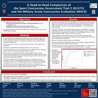
Catheterization and Cardiovascular Interventions 00:000–000 (2013) Original Studies Head-to-Head Comparison of Sirolimus-Eluting Stent Versus Bare Metal Stent Evaluation of the Coronary Endothelial Dysfunction in the Same Patient Presenting with Multiple Coronary Artery Lesions: The CREDENTIAL study Alexandru Nicolae Mischie,1,2*MD, Marco Stefano Nazzaro,1PhD, MD, Rosario Fiorilli,1MD, Francesco De Felice,1MD, Carmine Musto,1MD, Pierpaolo Confessore,1MD, Antonio Parma,1MD, Carla Boschetti,1MD, and Roberto Violini,1MD, PhD, FESC Objectives: To assess the endothelial dysfunction (ED) after bare metal stents (BMS) and sirolimus eluting stents (SES) implantation in the same patient, overcoming the confounding role of individual variables. compared to BMS but causes more ED. ED is a potentially unsafe phenomenon, since it is the first step in the cascade of atherosclerosis. Studies showing more pronounced ED with drug eluting stents than BMS involved different series of patients, making the comparison difficult because endothelial function (EF) is responsive to many risk fac- tors. Methods: we designed a prospective comparison of 6 months post-deployment EF of SES versus BMS implanted in the same patient, but in different coronary seg- ments. Forty-eight lesions were randomly assigned on a 1:1 allocation using block siz- ing of 4 according to a computer-generated sequence (SAS System, Version 9.1) basis to treatment with SES or BMS. The EF was evaluated by measuring vessel diameter variation in the stented segment, before and after selective intracoronary infusion of acetylcholine (iiAch). Results: In eligible patients, the relative magnitudes of major vasoconstriction were 2.6, 2.9, 4.6, and 3.1 at 5 mm proximal and 5, 10 and 20 mm dis- tal to the stent edge. Overall, a 3.5-fold major distal vasoconstriction after iiAch of SES vs. BMS was calculated. Conclusions: in the same patients, but treating different cor- onary segments, SES implantation induces a higher rate of vasoconstriction compared to BMS. The increased vasoconstriction after iiAch is an indicator of ED. Background: SES reduce restenosis rate C 2013 Wiley V Periodicals, Inc. Key words: endothelial function; acetylcholine; endothelium *Correspondence to: Mischie Alexandru Nicolae, U.O. Cardiologia Interventistica, Ospedale S. Camillo, C.ne Gianicolense n. 87, 00152 Roma, Italy. E-mail: alexandru_mischie@yahoo.com 1U.O. Cardiologia Interventistica, Ospedale San Camillo, C.ne Gianicolense n. 87, 00152, Roma, Italy 2Department of Cardiology, “Bagdasar-Arseni” Emergency Hospital, 12 Berceni Street, 4th Sector, 041915, Bucharest, Romania Grant sponsor: European Cardiovascular Interventions (EAPCI). ClinicalTrial.gov number NCT01242306. Received 8 August 2012; Revision accepted 21 January 2013 Association of Percutaneous DOI: 10.1002/ccd.24844 Published online in Wiley Online Library (wileyonlinelibrary.com). Conflict of interest: Nothing to report. C 2013 Wiley Periodicals, Inc. V
2 Mischie et al. INTRODUCTION discretion and these stenoses were excluded from the analysis (Fig. 1—Study Protocol). Particular attention was paid to obtain the best angiographic result and to implant only one stent for each lesion. All patients received optimal medical therapy, including aspirin 75 mg/day and clopidogrel 75 mg/day for at least 6 months. All patients were asked to return for invasive evaluation at 6 month of follow-up (FU). Medications with poten- tial effects on vasomotor responses were discontinued 72 hr before the procedure, but short-acting nitrates were permitted 6 hr prior to the procedure. Baseline cor- onary angiograms were taken and patients without intra- stent or peri-stent restenosis were included in the pharmacological protocol study. The protocol included intracoronary infusion of 0.9% normal saline (2 ml for 1 min), followed by baseline coronary angiography. The endothelium-dependent vasomotor response was esti- mated after a 2 min super-selective intracoronary infu- sion of acetylcholine 10?5mol/l (iiAch) using a pump (Perfusor Compact B. Braun Melsungen, Germany) and a coronary micro-catheter Corporation, Tokyo, Japan) positioned 3 cm above the proximal edge of the stent; after iiAch infusion, the micro-catheter was withdrawn and coronary angiograms were collected every 60 sec for 3 min (Fig. 2). The en- dothelial independent vasomotion was assessed 1 min after an intracoronary 200 mg bolus of nitroglycerin. Clinical status, heart rate, arterial pressure, and electro- cardiographic leads were continuously monitored. Sirolimus eluting stents (SES) reduce restenosis rate compared to bare metal stents (BMS) [1], but cause more pronounced endothelial dysfunction (ED) due to lack or delay of endothelization. The endothelium plays a critical role in vascular homeostasis by secreting sub- stances and influencing vascular inflammation and cell migration: lack or changes of this natural barrier for blood lipids and lipid deposition could trigger the atherogenesis process and could be associated with thrombogenicity. This potentially unsafe phenomenon [2], is the first step in the cascade of atherosclerosis [3,4], as well as a reliable predictor of future coronary events [5–7]. The studies showing more pronounced ED with SES than BMS involved different series of patients implanted with different types of stent, making the com- parisons difficult since endothelial function (EF) is responsive to many risk factors. To overcome the con- founding role of individual variables, we designed a prospective randomized comparison of 6-months post- deployment EF of SES versus BMS implanted in differ- ent coronary segments of the same patient. (FINECROSS, Terumo MATERIALS AND METHODS Study Design The study was conducted at the Interventional Cardi- ology Unit of the San Camillo-Forlanini Hospital, in Rome-Italy. Patients with stable angina pectoris, posi- tive stress test and indication to percutaneous coronary angioplasty (PCI) for at least 2 de novo >70% coronary stenoses were considered eligible. The two target lesions should have lengths >10 and <30 mm and comparable angiographic characteristics: differences measuring no more than 0.5 mm in reference vessel diameter and <50% in lesion length. Stents were implanted in differ- ent vessels or in the same vessels but in different ramifi- cations. The following variables were recorded for each patient: age, sex, body mass index, family history of heart disease, prior myocardial infarction, angina class, heart failure class, stroke or transient ischemic attack, chronic renal failure (defined as serum creatinine >2 mg/dl), diabetes mellitus, hypertension, smoking, dys- lipidemia, c-reactive protein and homocysteine levels. PCI was performed according to standard guidelines. The most severe of two lesions was randomly assigned to receive a SES (Cypher, Cordis Corporation, Miami Lakes, Florida) or a BMS (Coroflex Blue, B. Braun Melsungen, Germany) with allocation 1:1 using block sizing of 4 according to a computer-generated sequence (SAS System, Version 9.1). Consequently, a different stent was implanted on the second lesion. The treatment of further stenosis, if present, was left to the operator’s Exclusion Criteria Exclusion criteria were (a) clinical data: acute coronary syndrome in the last 3 months, severe risk factors for ED [uncontrolled diabetes mellitus (defined as HbA1C>9%), uncontrolled hypertension (systolic blood pressure >180 mm Hg), hypercholesterolemia (total cholesterol >240 mg/dl), persistent smoking]; any contraindication/intoler- ance to the use of aspirin, heparin, and/or clopidogrel; chronic renal failure requiring dialysis; severe left ven- tricular dysfunction (defined as an ejection fraction <35% by echocardiography); survival expectancy <1 year; (b) basal coronary angiographic findings: reference vessel diameter <2,5 mm, vasospasm, fresh thrombus, dissection, bifurcation/ostial lesions; (c) FU angiographic findings: restenosis (vessel diameter reduction >50%) or development of de novo significant stenosis (>70%). The study end-point was maximal coronary vasomo- tor response to iiAch. It was determined as a drug- induced percentage change in vessel diameter using baseline angiogram as reference. Eight points in the stent and peri-stent site and the proximal (10 mm proximal to the proximal stent edge) and distal (distal stent edge to 20 mm distally) average segment Catheterization and Cardiovascular Interventions DOI 10.1002/ccd. Published on behalf of The Society for Cardiovascular Angiography and Interventions (SCAI).
CREDENTIAL 3 Fig. 1. mus eluting stents; FU: follow-up. Study Protocol. AMI: acute myocardial infarction; BMS: bare metal stents; SES: siroli- diameters were analyzed (Fig. 2). The percent changes in vessel diameter after iiAch and nitrates were calcu- lated and compared between SES and BMS treated segments and baseline. Two orthogonal views with less foreshortening or without overlapping of side branches were selected and averaged for biplane assessment by twoexperts. End-diastolic images for each segment were chosen and quantitative coronary angiography (QCA) was performed using the CAAS II system (Pie Medical Imaging, Maastricht, The Netherlands). The contrast-filled tip catheter was used for calibration. In- dependent, masked reviewers performed the QCA measurements at baseline, after iiAch and nitrates. The independent predictors of ED were also investigated. from the reported cases. The Local Institutional Review Board approved the Protocol; all participants were provided with written, informed consent forms. The operators were aware of the assigned stent during PCI, but at angiographic FU and EF evaluation, staff was blinded to the allocation of stent type. A sample size has been calculated on the basis of previous reports [8–11] and we anticipated the occurrence of maximal vasoconstriction in response to iiAch meas- ured at 5 mm segments proximal and distal to the stent would be 40–60% and 0–20% respectively for SES versus BMS. Assuming a 0.05% alpha type error and 0.95 power, a total of 20 patients needed to be enrolled for a paired data study. To take into account potential losses to FU, we randomized 24 patients. Statistical analysis: baseline expressed as mean6SD and categorical variables as a number (n) and percentage (%). The univariate compari- sons between the continuous variables were performed characteristics are Conduct of the Clinical Study A masked, independent committee collected the end- points; an independent study monitor verified all data Catheterization and Cardiovascular Interventions DOI 10.1002/ccd. Published on behalf of The Society for Cardiovascular Angiography and Interventions (SCAI).
4 Mischie et al. Fig. 2. iAch: intracoronary Acetylcholine; PSE: proximal stent edge; DSE: distal stent edge; SES: sirolimus eluting stent; BMS: bare metal stent; *: P<0.05. Evaluation of endothelial function at 6-month follow-up (eight predefined points). using the Student’s t-test for paired and unpaired data. To identify the potential variables to enter into a multi- variate predictive model, we tested the correlation between clinical and angiographic variables. Multiple linear regression analysis was used to determine inde- pendent predictors of ED. Variables considered were: age, gender, diabetes, hypertension, current smoker, sta- tin, and angiotensin converting enzyme inhibitor/angio- tensin-receptor blocker used. A P value of <0.05 was considered statistically significant. SPSS statistical pro- gram (Chicago, IL) was used. TABLE I. Baseline Characteristics 70.867.1 1.8760.11 9 (64.2%) 4 (28.5%) 8 (57.1%) 13 (92.8%) 6 (42.8%) 5 (35.7%) Age (years) Body surface area (m2) Male sex Current smokers Positive family history of HD* Hypertension Prior myocardial infarction Diabetes mellitus type II Stable angina (CCS†) II III IV 7 (50%) 4 (28.5%) 3 (21.4%) *HD¼heart disease; †CCS¼Canadian Cardiovascular Society. Data are presented as numeric value (6 SD) and percentage (%). RESULTS During the study period, 540 patients were screened, 24 were enrolled and four patients were excluded: three (12.5%) refused the angiographic FU, one (4.1%) suf- fered acute coronary syndrome in a nonstented vessel. Angiographic FU was performed in 20 patients (83%), four (16.6%) had intrastent restenosis of the BMS, two (8.3%) showed significant progression of lesions in non-target vessels, and 14 patients entered in the final analysis. The flow diagram of the trial is provided in Fig. 1. Baseline clinical characteristics are presented in Table I. SES versus BMS had comparable angiographic and procedural characteristics (Table II). There were no differences between SES and BMS regarding mean stent length and mean stent diameter (19.2967.29 mm versus 16.162.9 mm, P¼0.25; 2.8660.39 mm versus 3.260.4 mm, P¼0.38, respectively). Among the BMS, there were ten direct stents with- out post-dilatation and four stent implantations with pre- and post-dilatations; among the SES there were nine direct stents without post-dilatation and five stent implantations with pre and post-dilatations. The mean interval from stent implantation to FU angiography was 180.4610.3 days. Blood sample results at FU are listed in Table III. The percentage variation in vessel diameters after iiAch in the eight predefined points and two segment mean diameters for SES and BMS are shown in Figs. 2 and 3. Catheterization and Cardiovascular Interventions DOI 10.1002/ccd. Published on behalf of The Society for Cardiovascular Angiography and Interventions (SCAI).
CREDENTIAL 5 TABLE II. Angiography Characteristics The relative magnitudes of major vasoconstriction for SES versus BMS were 2.6 (P¼0.04), 2.9 (P¼0.03), 4.6 (P¼0.001) and 3.1 (P¼0.002) at 5 mm proximal and 5, 10, and 20 mm distal to the stent edge. Overall, a 3.5-fold major distal vasoconstriction after iiAch of SES versus BMS was calculated as an average of the three predefined distal diameters (coronary angiography of patient n. 11 in Fig. 4 after iiAch and nitrates). In univariate analysis, the independent significant predictors that correlated with increased ED were SES, diabetes, hypertension, low High Density Lipoprotein levels, presence of atherosclerosis distal to SES im- plantation site, age, increased C-Reactive Protein levels and prior myocardial infarction. After intracoronary nitrates were administered, there were no statistically significant differences in vessel diameters between SES versus BMS for any of the evaluated segments. No differences were recorded in the mean arterial blood pressure during the iiAch compared to the base- line. Two patients had an episode of temporary asysto- lia (less than 5 sec) that recovered spontaneously. Intraobserver and interobserver variability for quantita- tive measurements of coronary angiography in the same recordings of 15 randomly selected vessels were 0.06260.04 mm and 0.02160.03 mm, respectively. BMS (n¼14) SES (n¼14) P value Vessel location Left anterior descending Left circumflex Right Ramus intermedius ACC/AHA lesion class A B1 B2 Angiographic measures Lesion length (mm) Reference vessel diameter (mm) Minimal lumen diameter (mm) Stenosis (% of lumen diameter) Stent diameter (mm) Stent length/lesion (mm) Maximal pressure/lesion (atm) 4 (28.5%) 5 (35.7%) 5 (35.7%) 0 5 (35.7%) 3 (21.4%) 4 (28.5%) 2 (14.2%) ns ns ns 0.05 7 (50%) 4 (28.5%) 3 (21.4%) 6 (42.8%) 4 (28.5%) 4 (28.5%) ns ns 0.05 15.965.4 3.160.60 0.8560.64 7865.6 3.260.4 16.162.9 15.7464.6 16.667.2 2.8560.89 0.8860.56 80610.4 2.8660.39 19.267.2 16.264.2 ns ns 0.05 ns ns ns 0.05 Data are presented as numeric value (6 SD) and percentage (%). TABLE III. Blood Samples Results at 6-Month Follow-Up 0.0960.04 0.960.2 6.060.6 9.962.5 13.561.3 155.9629.7 78.8619.1 45.9613.8 112.1664.2 302.6645.9 C-Reactive Protein (mg/dl) Creatinin (mg/dl) HbA1C (%) Homocysteine (mm/l) Hemoglobin (g/dl) Total cholesterol (mg/dl) Low density lipoprotein (mg/dl) High density lipoprotein (mg/dl) Triglyceride (mg/dl) Fibrinogen (mg/dl) DISCUSSION This study shows that in the same group of patients, the 6-month FU endothelial Data are presented as numeric values6SD. dependent coronary Fig. 3. nary arteries, evaluated during intracoronary infusion of Acetylcholine (6-month follow-up). Prox.: proximal; Dist.: distal; *: P<0.05; SD: standard deviation; SES: sirolimus eluting stent; BMS: bare metal stent. [Color figure can be viewed in the online issue, which is available at wileyonlinelibrary.com.] Endothelial function after BMS and SES implantation in the same patient but different coro- Catheterization and Cardiovascular Interventions DOI 10.1002/ccd. Published on behalf of The Society for Cardiovascular Angiography and Interventions (SCAI).
6 Mischie et al. tive intracoronary Ach infusion (1025mol/l); D: recovery recov- ery after 200 mg bolus of nitroglycerin. SES: sirolimus eluting stent; BMS: bare metal stent; Ach: Acetylcholine. [Color figure can be viewed in the online issue, which is available at wileyonlinelibrary.com.] Patient number 11.•5SES: 2.75 3 23.00 mm on proxi- Fig. 4. mal left anterior descendent. A: selective intracoronary Ach infusion (1025 mol/l), notice (arrows); B: recovery after 200 mg bolus of nitroglycerin. •5BMS: 3.00 x 19.00 mm on right coronary artery. C: selec- the distal vasoconstriction vasomotion is significantly impaired after SES com- pared to BMS implantation, especially distal to the stent. Instead, no differences exist between the two types of stent in regards to endothelium independent coronary vasomotion. This data supports that SES, rather than BMS implantation is associated with coro- nary ED [9]. Previous studies have demonstrated more pronounced ED after SES versus BMS, but the com- parisons have been assessed in different patients with different risks factors [10–12]. Because EF is a com- plex process influenced by a number of pathophysio- logical mechanisms (ethnicity, hypertension, diabetes, smoking, hypercolesterolemia etc.), a considerable vari- ability exists in the healing process after stent implan- tation. In our study, both types of stent were randomly implanted in two comparable coronary lesions within the same patient. This unique study design actually adjusts the comparison for all variables and definitively assesses the impact of the type of stent on EF. A methodological issue is the protocol of study of coronary EF. The QCA response after iiAch is currently the most utilized inva- sive tool of investigation of coronary vasomotion [10,11]. The drug acts as a potent vasodilator in nor- mal coronary vessels by promoting the release of endo- thelial nitric oxide, but in damaged vessels it can cause abnormal vasoconstriction via receptors localized in the smooth muscle cells [13]. IiAch provocation test is a sensitive and safe test, biased by the lack of stand- ardization because a number of different protocols— fixed versus progressive doses, low versus moderate versus high doses, manual versus pump injections, non- selective (a guiding catheter in the coronary ostium) versus superselective (a microcatheter in the target ves- sel)—all produce variable drug concentrations, poten- tially contributing to different results of EF after SES or BMS implantation. As an original finding of the study, we aimed to obtain the best control of drug evaluation of coronary Catheterization and Cardiovascular Interventions DOI 10.1002/ccd. Published on behalf of The Society for Cardiovascular Angiography and Interventions (SCAI).
CREDENTIAL 7 concentration by infusing iiAch “super-selectively” into the target vessel. We chose a fixed, single dose (10?5mol/l) of the drug, which provides a moderate, consistent vasocostrictive stimulus and ensures a very low rate of potential, serious complications. Our results are in line with previous studies where similar dose of iiAch were used [8,10–12]. Our data addresses the open issues of potential adverse biological effects of SES: coronary endothelium-dependent vasomotion is severely impaired after SES implantation, while it is virtually unaffected after BMS implantation [9,11]. Over 20% vasoconstriction after iiAch has been con- sidered a reliable sign of ED [14]. Many studies have documented the association between ED and serious cardiovascular events [15,16]. It is common experience to detect a higher incidence of restenosis at the edge of the SES, which could be explained by the presence of ED. This phenomenon is consistent with the results of our study, that shows major ED in the same area most affected by restenosis of the SES. Several cases of diffuse coronary spasm after SES implantation have been reported [17,18] and ED is implicated in the increased incidence of very late stent thrombosis with first generation of drug eluting stents (DES), especially after discontinuation of dual antiplatelet therapy. These adverse effects could offset the potential benefits of SES. We have to define the duration of SES-induced- ED and evaluate if we can counterbalance its negative effects. In agreement with a Consensus for Preclinical stent evaluation that recommended evaluation of EF as a valuable ancillary tool for differentiating the long- term performance of DES [19], questions raised by this work highlight the need for additional investigations. Compared to extensive use of DES in the current treatment of ischemic heart disease, EF after SES-PCI focuses on a lingering issue. For the millions of patients in whom SES have already been deployed, aggressive efforts to improve general EF is a “gray” area which is not given sufficient consideration. A potential clinical use of our results highlights the bio- logical effects of SES in terms of EF identifying the patients with increased endothelial sensitivity that require special targeted medical or interventional treatment. attempted due to the small sample size. A longer FU with a second invasive evaluation of EF at 1 year would have allowed an evaluation of the recovery timeframe, if present. SES were chosen because, at the time, they were the most studied and most implanted stents in the real world, and, despite the fact that they are no longer used today, they have been implanted in millions of patients. Finally, a similar evaluation of EF of second-gen-DES would be equally interesting. CONCLUSIONS In the same patients, but treating different coronary segments, SES implantation induces a higher rate of ED compared to BMS. The increased vasoconstriction after iiAch is an indicator of ED. We calculated: a 3.5- fold vasoconstriction of SES vs. BMS for the three punctual distal diameters and a 1.9-fold vasoconstric- tion of SES vs. BMS for the distal segment average di- ameter. ClinicalTrial.gov number NCT01242306. REFERENCES 1. Morice MC, Serruys PW, Sousa JE, Fajadet J, BanHayashiE, Perin M, Colombo A, Schuler G, Barragan P, Guagliumi G, Moln? ar F, Falotico R. A randomized comparison of a sirolimus- eluting stent with a standard stent for coronary revascularization. N Engl J Med 2002;346:1773–1780. 2. Suzuki T, Kopia G, Hayashi S, Bailey LR, Llanos G, Wilensky R, Klugherz BD, Papandreou G, Narayan P, Leon MB, Yeung AC, Tio F, Tsao PS, Falotico R, Carter AJ. Stent-based delivery of sirolimus reduces neointimal formation in a porcine coronary model. Circulation 2001;104:1188–1193. 3. Drexler H, Zeiher AM. Progression of coronary endothelial dys- function in man and its potential clinical significance. Basic Res Cardiol 1991;86(Suppl 2):223–232. 4. Nobuyoshi M, Tanaka M, Nosaka H, Kimura T, Yokoi H, Hamasaki N, Kim K, Shindo T, Kimura K. Progression of coro- nary atherosclerosis: Is coronary spasm related to progression? J Am Coll Cardiol 1991;18:904–910. 5. Suwaidi JA, Hamasaki S, Higano ST, Nishimura RA, Holmes DR Jr, Lerman A. Long-term follow-up of patients with mild coronary artery disease and endothelial dysfunction. Circulation 2000;101:948–954. 6. Halcox JP, Schenke WH, Zalos G, Mincemoyer R, Prasad A, Waclawiw MA, Nour KR, Quyyumi AA. Prognostic value of coronary vascular endothelial 2002;106:653–658. 7. Heitzer T, Schlinizing T, Krohn K, Meinertz T, Munzel T. En- dothelial dysfunction, oxidative stress, and risk of cardiovascular events in patients with coronary artery disease. Circulation 2001;104:2673–2678. 8. Kim JW, Seo HS, Park JH, et al. A Prospective, Randomized, 6 months comparison of the coronary vasomotor response associ- ated with a zotarolimus versus a sirolimus eluting stent: Diferen- tial recovery of coronary endothelial dysfunction. J Am Coll Cardiol 2009;53;1653–1659. 9. Togni M, Windecker S, Cocchia R, Wenaweser P, Cook S, Bill- inger M, Meier B, Hess OM. Sirolimus-eluting stents associated dysfunction. Circulation Study Limitations The number of enrolled patients was limited due to the high rate of drop-out at the angiographic FU, so the final power analysis of the study changed from 0.95 to 0.85 (maintaining an 0.05% alpha type error). At the FU angiogram, the stent type was incompletely masked because a BMS is clearly thinner than the SES. The multiple linear regression analysis was not Catheterization and Cardiovascular Interventions DOI 10.1002/ccd. Published on behalf of The Society for Cardiovascular Angiography and Interventions (SCAI).
8 Mischie et al. with paradoxic coronary vasoconstriction. J Am Coll Cardiol 2005;46:231–6. 10. Hofma SH, van der Giessen WJ, van Dalen BM, Lemos PA, McFadden EP, Sianos G, Ligthart JM, van Essen D, de Feyter PJ, Serruys PW. Indication of long-term endothelial dysfunction after sirolimus-eluting stent 2006;27:166–170. 11. Fuke S, Maekawa K, Kawamoto K, Saito H, Sato T, Hioka T, Ohe T. Impaired endothelial vasomotor function after sirolimus- eluting stent implantation. Circ J 2007;71:220–225. 12. Caramori PRA, Lima VC, Seidelin PH, Newton GE, Parker JD, Adelman AG. Long-term endothelial dysfunction after coronary artery stenting. J Am Coll Cardiol 1999;34:1675–1679. 13. Farouque HMO, Meredith IT. The assessment of endothelial function in humans. Coron Artery Dis 2001;12:445–454. 14. Maseri A, Davies G, Hackett D, Kaski JC. Coronary artery spasm and vasoconstriction. The case for a distinction. Circula- tion 1990;81:1983–1991. 15. Schachinger V, Britten MB, Zeiher AM. Prognostic impact of coronary vasodilator dysfunction on adverse long-term outcome of coronary heart disease. Circulation 2000;101:1899–1906. 16. Bonetti PO, Lerman LO, Lerman A. Endhotelial dysfunction: A marker of atherosclerotic risk. Aterioscler Thromb Vasc Biol 2003;23:168–175. 17. Brott BC, Anayiotos AS, Chapman GD, Anderson PG, Hillegass WB. Severe, diffuse coronary artery spasm after drug eluting- stent placement. Invasive Cardiol 2006;18:584–592. 18. Maekawa K, Kawamoto K, Fuke S, Yoshioka R, Saito H, Sato T, Hoioka T. Images in cardiovascular medicine. Severe endo- thelial dysfunction after sirolimus eluting stent implantation. Circulation 2006;113:e850–e851. 19. Schwartz RS, Edelman E, Virmani R, Carter A, Granada JF, Kaluza GL, Chronos NAF, Robinson KA, Waksman R, Weinberger J, Wilson GJ, Wilensky RL. Drug-eluting stent in preclinical studies. Updated consensus recommendations for preclinical evaluation. Circ Cardiovasc Interv 2008;1:143–153. implantation. Eur Heart J Catheterization and Cardiovascular Interventions DOI 10.1002/ccd. Published on behalf of The Society for Cardiovascular Angiography and Interventions (SCAI).