Download
1 / 19
190 likes | 408 Vues
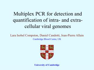
Multiplex PCR for detection and quantification of intra- and extra-cellular viral genomes. Lara Isobel Compston, Daniel Candotti, Jean-Pierre Allain Cambridge Blood Centre, UK. University of Cambridge. Introduction.

E N D
Multiplex PCR for detection and quantification of intra- and extra-cellular viral genomes Lara Isobel Compston, Daniel Candotti, Jean-Pierre Allain Cambridge Blood Centre, UK University of Cambridge
Introduction • Interaction between viruses with the host is dependant on the interplay between factors related to both the virus and the host • Target cells can be damaged either directly by the virus or by the immune response initiated, and an equilibrium needs to be reached between them • Several viral survival strategies are the result of the virus/host interplay : 1) Clinical recovery due to successful development of humoral and cellular immune responses. 2) Latent viral infection (transient escape of immune response in primary infection and reactivation) 3) Establisment of chronic viral infection with partially effective immune response.
Lymph nodes CMV, HHV-8, EBV, HIV • Clinical symptoms • Clinical recovery • By immune defenses • Antibodies • White cells • No detectable virus in circulation Liver HBV, HAV, HCV Bone marrow HEV B19 New model of viral infection Recent evidence has emerged that, in common with latent viruses, after recovered infections, common viruses are not eliminated from the host but contained efficiently, persisting in sanctuaries were they escape the immune response. Acute infection Sanctuaries: biological portfolio Recipients of TX or organ Immunocompetent Maintain viral control Undetectable in blood Immunodeficient Age, chemo; transplant of BM or organs; HIV - Increased susceptibility to external pathogens - Reactivation of past infections ( viral load) - Severe symptoms
Aims of the study • It was hypothesised that the generality of reactivation of common latent and persistent viral genomes may constitute an indicator of the overall immune status of the host. • The objective was to systematically detect and quantify a panel of common viruses in persons who are either immunocompetent or present with varying degrees of immunodeficiency ranging from mild to severe.
Multiplex real-time PCR B19 / HBV/ HHV-8 EBV/CMV/ VZV GBV-C / HAV PCR signal for specific viruses No Yes Sample negative for specific virus Single virus qPCR Viral load quantification Final viral load result (>50 copies) ≤ 50 copies undetectable Nested-PCR No Yes Confirmation of ≤ 50 copies Sample negative for specific virus Screening algorithm
Development of Standards for qPCR • NIBSC standards: • WHO international standard for Hepatitis B virus 97/746 • WHO international standard for Hepatitis A virus 00/560 • Plasmid standards: • Constructed for the following targets: • EBV and HHV-8 (cell culture) • CMV (clinical sample) • VZV (oligonucleotide construct of target region) • B19 (received as a gift) • PCR amplicons of target regions • Plasmids purified and quantified by UV spectroscopy, plasmid concentration used to derive copy number by standard conversion for qPCR • Clinical standards: • A high titre clinical sample of GBV-C serially diluted and used as a standard • Unknown initial viral load, therefore dilution giving 100% L.O.D designated as 10 AU (arbitrary units)
HBV Hepatitis A virus Dynamic range with NIBSC standards 95% C.I. values shown
NIBSC standards: • WHO international standard for Hepatitis B virus 97/746 • WHO international standard for Hepatitis A virus 00/560 • Plasmid standards: • Constructed for the following targets: • EBV and HHV-8 (cell culture) • CMV (clinical sample) • VZV (oligonucleotide construct of target region) • B19 (received as a gift) • PCR amplicons of PCR target region • Plasmids purified and quantified by UV spectroscopy, plasmid concentration used to derive copy number by standard conversion for qPCR • Clinical standards: • A high tire clinical sample of GBV-C serially diluted and used as a standard • Unknown initial viral load, therefore dilution giving 100% L.O.D designated as 10 AU (arbitrary units)
Dynamic range with plasmid standards 95% C.I. values shown CMV VZV HHV-8 EBV
B19 50 % Limit of detection Dynamic range with plasmid standards 95% C.I. values shown
NIBSC standards: • WHO international standard for Hepatitis B virus 97/746 • WHO international standard for Hepatitis A virus 00/560 • Plasmid standards: • Constructed for the following targets: • EBV and HHV-8 (cell culture) • CMV (clinical sample) • VZV (oligonucleotide construct of target region) • B19 (received as a gift) • PCR amplicons of PCR target regions • Plasmids purified and quantified by UV spectroscopy, Plasmid concentration used to derive copy number by standard conversion for qPCR • Clinical standards: • A high titre clinical sample of GBV-C serially diluted and used as a standard • Unknown initial viral load, therefore dilution giving 100% L.O.D designated as 10 AU (arbitrary units)
Dynamic range with clinical standards 95% C.I. values shown GBV-C
Multiplex assays for DNA viruses relevant in Africa B19 singleplex = limit of detection; 50 IU Hepatitis B singleplex = limit of detection; 50 IU Rsq 0.995 Rsq 0.992 Ct of 50 IU = 35.02 Ct of 50 IU = 34.95 Multiplex: limit of detection; B19 500 IU, HBV 50 IU, HHV-8 10 copies HHV-8 singleplex = limit of detection;10 copies B19 Rsq 0.939 Ct of 500 IU: 34.59 Rsq 0.977 HBV Rsq 0.996 Ct of 50 IU: 35.43 HHV-8 Rsq 0.953 Ct of 10 copies: 33.67 Ct of 10 copies = 34.04
Multiplex assay for DNA viruses relevant worldwide EBV Singleplex: limit of detection; 214 copies CMV Singleplex: limit of detection; 350 copies Rsq 0.995 Rsq 0.941 Ct of 214 copies = 35.28 Ct of 350 copies = 34.46 Multiplex: limit of detection; VZV 217 copies, EBV 214 copies, CMV 350 copies VZV Singleplex: limit of detection; 217 copies EBV Rsq 0.995 Ct of 214 copies = 34.53 Rsq: 0.981 VZV Rsq 0.997 Ct of 217 copies = 39.24 CMV Rsq 0.998 Ct of 350 copies = 37.4 Ct of 217 copies = 36.59
RNA virus Duplex Hepatitis A singleplex: limit of detection; 40 IU Rsq 0.853 Duplex: Limit of detection; GBV-C 10 AU, HAV 40 IU HAV Rsq 0.636 Ct of 40 IU = 35.79 Ct of 40 IU = 37.59 GBV-C Rsq 0.880 Ct of 100 AU = 37.0 GBV-C singleplex: limit of detection; 10 AU Rsq 0.942 Ct of 100 AU = 37.42
1.0x10 07 1000000 100000 10000 1000 100 10 1 0 CMV VZV EBV HHV-8 HBV B19 GBV-C HAV qPCR results in Ghanaian blood donors Limit of detection
97.1 97.1 100 93.4 90 80 72.4 70 58.4 60 % Positive 46.3 50 Viraemia Ab 40 22.8 30 13.4 20 3.9 10 0.49 0 0.99 0.49 0.49 0.49 0 0 VZV CMV EBV HHV-8 HBV B19 GBV-C HAV Virus Background serology and viraemia in Ghanaian blood donors
Summary • A range of different types of standards where utilised for qPCR, which had similar dynamic ranges, repeatability and reproducibly • NIBSC standards • Plasmid standards • Clinical standards • Triplex assays where developed and optimised to match the sensitivity of the singleplex PCR • This was achieved, except for B19 ( one-log decrease in sensitivity) • The HAV assay could not be utilised as a quantitative assay despite extensive optimisation • The Ghanaian blood donor population, while having high seroprevalence for each virus examined indicating previous exposure, had only very low frequency and level of viraemia • No viraemia was detected with HHV-8, VZV and HAV despite high background seroprevalence • Against this background studies of reactivation in various situations of immunodeficiency can now be conducted
Acknowledgements • Division of Transfusion Medicine (University of Cambridge) • Jean-Pierre Allain • Daniel Candotti • Komfo Anokye Teaching Hospital (Kumasi, Ghana) • Ohene Opare-Sem • Francis Sarkodie • Laboratoire de Virologie (Hôpital Armand Trousseau) • A. Garbarg-Chenon