Download
1 / 54
570 likes | 827 Vues
The Blood. VNSG 1420 Anatomy & Physiology Chapter 12. Blood. Fundamental to maintaining homeostasis Pumped by the heart through a closed system of blood vessels Classified as a connective tissue cells make up nearly half of blood Viscous (thick) fluid

E N D
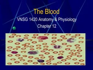
The Blood VNSG 1420 Anatomy & Physiology Chapter 12
Blood • Fundamental to maintaining homeostasis • Pumped by the heart through a closed system of blood vessels • Classified as a connective tissue • cells make up nearly half of blood • Viscous (thick) fluid • color depends based on oxygen content • 5 liters in average adult male • 8% of total body weight
Functions of Blood • Transportation • oxygen, carbon dioxide, nutrients, minerals, vitamins, hormones & wastes • Regulation • of pH, fluid balance & body temperature • Protection • against foreign organisms & blood loss
Transportation • Gases • Oxygen & carbon dioxide • Nutrients and other needed substances • Nutrients, electrolytes (salts) & vitamins • Waste • Water, acid, electrolytes, urea, pigments, hormones, drugs, carbon dioxide • Hormones • from their sites of origin • to the organs they affect
Regulation • pH • Buffers in blood help to keep body fluid pH at about 7.4 • Fluid balance • Regulates amount of fluid in tissues by maintaining proper osmotic pressure • Heat • Transports heat generated in muscles to other parts of the body, regulating body temperature
Protection • Disease • Important defense against disease • Carries cells & antibodies of the immune system that protect against pathogens • Blood loss • Contains factors that protect against blood loss from the site of an injury
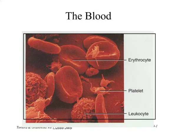
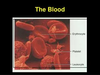
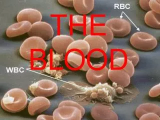
Blood Constituents • Plasma - liquid portion of blood • Formed elements - cells & fragments • Erythrocytes - red blood cells - transport oxygen • Leukocytes - white blood cells - protect against infection • Platelets - thrombocytes - cell fragments that aid in blood clotting
Blood Plasma • 55% of total blood volume • 91% water • 8% Proteins • albumin, clotting factors, antibodies & complements • Remaining 1% = • Nutrients - carbohydrates, lipids & amino acids • Electrolytes - minerals • Wastes, hormones, vitamins & drugs, dissolved gases
Plasma Proteins • Albumin • most abundant plasma protein, manufactured in liver, important in maintaining blood osmotic pressure • Clotting factors • needed for blood coagulation, produced in liver • Antibodies • combat infection • Complement • system of enzymes needed to help fight pathogens
Plasma Nutrients • Glucose - Main carbohydrate in plasma • Stored in liver and skeletal muscles as glycogen, released for energy • Absorbed by capillaries of intestine after digestion • Amino acids - products of protein digestion • Absorbed by intestine capillaries also • Lipids – fats, cholesterol & lipoproteins
Formed Elements • Erythrocytes - red blood cells (RBCs) • transport oxygen • Leukocytes - white blood cells (WBCs) • protect against infection • Platelets (thrombocytes) • needed for clotting of blood • All formed elements (blood cells) form from hematopoietic stem cells in red bone marrow (ancestors of all blood cells) See Table 10-2, p. 313
Erythrocytes • Red blood cells (RBCs) – biconcave (disk) shaped • Carry oxygen bound to hemoglobin • Most numerous of blood cells - 4.5 to 5 million per cubic millimeter of blood • Hemoglobin - protein that contains iron • Blood color determined by oxygen content • from lungs - bright red = oxygenated • to lungs - darker red = deoxygenated • Mature red cells have no nucleus & cannot divide, must be replaced constantly
More on Erythrocytes . . . • Hemoglobin able to carry hydrogen ions (acidic) • Acts as buffer to keep pH of 7.4 (acid-base balance) • Transport carbon dioxide from tissues to lungs for elimination • Carbon monoxide blocks ability of hemoglobin to carry oxygen • Erythropotein - hormone from kidney • stimulates production of red blood cells in response to decreased oxygen supply
Leukocytes • White blood cells (WBCs) • 5,000 to 10,000 per cubic millimeter of blood (round shape) • Colorless • Contain prominent nuclei • Different types identified by size, shape of nucleus & appearance or lack of granules in cytoplasm when stained (Wright’s stain) • % of each cell type useful in diagnosis
Granulocytes/ Agranulocytes • GRANULOCYTES • Neutrophil • Polymorphs, polys, segs • Eosinophil • Basophil • AGRANULOCYTES • Lymphocytes • Monocyte
Granular Leukocytes / Granulocytes • Granules visible when stained • Neutrophils - most numerous of WBCs • Active in fighting infections • Eosinophils • increase during allergic reactions & parasite infestation • Basophils • increase during allergic reactions & inflammatory reactions
More on Neutrophils • Various shaped nuclei • Polymorps or polys • Segmented nucleus - segs • Polymorphonuclear neutrophils -PMN • Immature nuclei - thick curved bands • Increase in band cells (stab or staff cells) is sign of infection & active neutrophil production
Agranular Leukocytes / Agranulocytes • Lack easily visible granules • Lymphocytes • Active in immunity • Second most numerous of WBCs • Monocytes • Largest in size • Function as phagocytes
Function of Leukocytes • Destruction of pathogens • Rid body of foreign materials - cell debris & pathogens • Engage in phagocytosis • Engulfing of pathogens [Neutrophils & monocytes leave blood vessels when pathogens enter tissues] • Travel by ameboid motion to area Pus = mixture of live & dead bacteria & live & dead leukocytes Abscess = pus localized in an area
More on Leukocyte Function . . . • Monocytes enter tissues, enlarge & mature into macrophages • Macrophages • active in disposal of pathogens or foreign material • Some lymphocytes become plasma cells • Plasma cells • lymphocytes active in producing circulating antibodies needed for immunity
Platelets / Thrombocytes • Smallest of formed elements - cell fragments from megakarocytes (large bone marrow cells), not cells • No nuclei or DNA • 150,000 to 450,000 per cubic millimeter • Essential in blood coagulation (clotting) • In blood vessel injury • platelets stick together to form plug that seals wound • platelets then release chemicals to form clot to stop blood loss
Hemostasis • Process that prevents blood loss from blood vessel rupture or injury • Hemostasis events • Contraction of blood vessels • Vasoconstrictionreducies diameter of vessel reducing blood flow • Formation of platelet plug • Formation of blood clot
Blood Clotting / Coagulation • Balance of regulating inactive compounds in blood stream: • Procoagulants - promote blood clotting • Activate with injury to form clot • Anticoagulants - prevent blood clotting • Prevail in normal conditions • 12 clotting factors involved in clotting process
Clotting Factors • Factors I - XII - blood clotting factors • Final steps in clotting = conversion of fibrinogen to fibrin • Substance from damaged tissue form prothrombinase – which converts prothrombin to thrombin • Thrombin converts fibrinogen(plasma protein) to solid threads of fibrin • Network of fibrin threads form clot • Serum - fluid that remains after blood clots PLASMA = SERUM + CLOTTING FACTORS
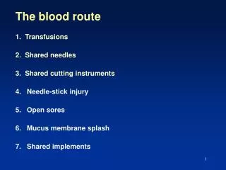
Blood Types • Blood transfusions • transfer of blood from 1 person to another - given for hemorrhage (excessive bleeding) or disease - hemorrhage results in oxygen & nutrients to cells • Incompatible transfusions • cause destruction of donor red cells (hemolysis) & transfusion reactions • Antigens – proteins on surface of RBCs that react with antibodies – to cause incompatibility
ABO Blood Type Group • Blood type describes type of antigen present on red blood cells • Type A has A antigenonly • Type B has B antigen only • Type AB has A & B antigens • Type O has no antigen
Testing For Blood Type • Tested by mixing blood sample with anti-sera to different antigens • Blood sera containing antibodies to A or B antigen prepared • Sera applied to blood sample • Agglutination occurs – clumping together of red cells when mixed with matching anti-serum [Type A reacts with Anti-A only; Type B reacts with Anti-B only; Type AB reacts with both; Type O reacts with NEITHER] See Table 10-3 page 319 & Figure 10-8, page 321
Blood Compatibility • Blood type determined by heredity • In an emergency, Type O blood can be given to any ABO type • Type O is universal donor • Blood Type AB can receive from any ABO type • Safest to give blood of same type in normal conditions
THE Rh Factor Antigens – proteins on the surface of red cells Antibody – substance produced in response to a specific antigen • Rh factor / D antigen – red cell antigen group • Rh positive (+) blood has Rh antigen • Rh negative(-) blood has no Rh antigen • Rh positive blood given to Rh negative person will produce antibody to Rh antigens
Rh Incompatibility Potential problem in pregnancy • Rh (-) mother can develop antibodies to RH protein of Rh (+) fetus • RBCs can enter mother’s circulation during pregnancy & childbirth & cause immune response • In subsequent pregnancy with Rh (+) fetus, anti-Rh antibodies from mother can pass to fetus causing fetal RBC destruction
Hemolytic Disease of Newborn (HDN) • Rhogam • Rh (D) immune globulin (antibody) is given to Rh (-) mother during pregnancy & after delivery to clear antigens from her circulation & prevent immune system response • Destruction of RBCs of Rh (+) fetus by anti-Rh antibodies of sensitized Rh (-) mother • Infant with Hemolytic Disease of Newborn (HDN) can receive lifesaving RH negative replacement blood transfusion
Use of Blood & Blood Components • Blood can be stored up to 35 days by blood bank • Anti clotting solution, • Expiration date • Important to keep extra Type O (universal donor) • Whole blood transfusions - used only to replace large volume blood losses • Massive hemorrhage from serious mechanical injury • Internal bleeding, as with bleeding ulcers • During or after surgery • Blood replacement for Hemolytic Disease of Newborn
More Types of Transfusions . . . • Autologous – donated for a person’s own use • Elective surgery • Blood component transfusions – formed elements are separated by centrifugation • Plasma components & expanders • Hemapheresis – desired elements kept, remainder returned to donor • Plasmapheresis – plasma kept, formed elements returned to donor
Use of Plasma • Replace blood volume • Prevent or treat circulatory failure (shock) • Because no RBCs, no incompatibility problems • Further separation • Protein fractions (to treat plasma protein deficiency) • Serum albumin • Immune serum • Clotting factors (cryoprecipitate obtained by freezing, contains clotting factors)- replacement use • Gamma globulin (contain antibodies - used for replacement)
Blood Studies • Standard part of routine physical exams • Hematocrit & Hemoglobin • Blood cell studies • Blood slide (smear) • Blood chemistry tests • Coagulation studies • Many tests done by machines
Hematocrit • Measures volume percentage of packed red blood cells in whole blood • # of mls of RBCs per 100mls of whole blood • Blood is spun in centrifuge to separate cellular elements from plasma • Normal hematocrit (Hct) ranges • Adult males - 42-54 • Adult female - 36-46 • Below normal levels of RBCs signify anemia See Figure 10-1, p. 311
Hemoglobin Tests • Measures grams of hemoglobin per 100 mls of whole blood • Hemoglobin needed for oxygen delivery to tissues • Color of blood is compared to a color scale to measure hemoglobin released from red cells • Normal hemoglobin (Hgb) ranges • Adult males - 14-17 grams • Adult females - 12-15 grams • Below normal levels of hemoglobin signify anemia
More on Hemoglobin Tests . . . • Electropheresis • Process that measures normal & abnormal types of hemoglobin • Electric current is passed through liquid that contains hemoglobin • Useful in diagnosis sickle cell anemia
Blood Cell Counts • RBC & WBC • Automated methods or visual counts under a microscope • Platelets counts (automated) • Normal counts • RBC = 4.5 to 5.5 million/cubic millimeter (mm) • WBC = 5000 to10,000/cubic mm • Platelets = 150,000 to 450,000/cubic mm
Blood Cell Count Changes • Increased RBC count is polycythemia • Decreased RBC count is anemia • Increased WBC count is leukocytosis • Decreased WBC count is leukopenia
Blood Smear Slide • Complete blood count (CBC) • Includes Hgb & Hct (H&H), blood cell counts & stained blood slide for differential cell counts • Blood smear - blood drop spread thinly & evenly over glass slide & stained with Wright’s stain • Red cells & platelets studied for abnormalities • Also look for parasites • % of different WBCs –differential white cellcount( add up to 100%)
Blood Chemistry Tests • Tests on blood serum typically by machine • Electrolytes - sodium, potassium, chloride & bicarbonate • Blood glucose – glucose/sugar in blood • Blood urea nitrogen (BUN) & creatinine - nitrogen waste products • Enzymes - CK, LDH, & others - can indicate tissue damage; an excess of Alkaline Phosphatase can indicate liver disorder or cancer