Medical mycoses
Medical mycoses. cutaneus subcutaneus systemic opportunistic. Intact skin and mucosal surfaces serve as barriers to infection by mycotic agents. Fatty acid,pH,epithelial turnover of the skin Transferrin to restrict the growth of several fungi.

Medical mycoses
E N D
Presentation Transcript
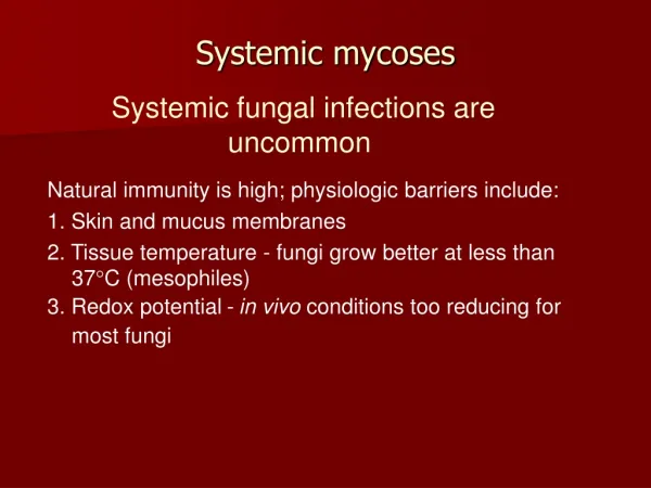
Medical mycoses cutaneus subcutaneus systemic opportunistic
Intact skin and mucosal surfaces serve as barriers to infection by mycotic agents. • Fatty acid,pH,epithelial turnover of the skin • Transferrin to restrict the growth of several fungi. • Superficial fungi dont elicit a cellular response from the host
Etiology • Mallassezia furfur • Pityriasis Versicolor • Lipophillic organism • Lesion: hypopigmented area contain budding yeast cells and hyphae • Asymptomatic • Diagnosis: KOH preparations(skin scrapping)
Dermatophytes • Dermatophytoses(Tinea,ringworm) • Infect keratinized structures • Skin,hair and nail • Invasive properties • May evoke inflamatory reaction • Three genera: - Epidermophyton - Trichophyton - Microsporum
Anthropophillic • Zoophillic • Geophillic Tinea chronic infection (warm and humid areas of the body) Typical lesion : inflame circular border containing papules & vesicles surrounding a clear area.
Broken hairs and thickened broken nails Trichophyton • tinea capitis in children • Endothrix infections • “favus” tinea capitis in which crusts on the scalp.
Dermatophytid (“id”) reactions - vesicles on the fingers - response to circulating fungal antigens - the lesion do not contain hyphae • Skin test with fungal extracts,eg, trichophytin
The normal skin is generally resistant to invasion by dermatophytes • In conditions of excessive moisture,can invade keratinized structures.
Laboratory diagnosis • Microscopic KOH preparation scrappings of skin or nail show hyphae 2. Cultures on Sabouraud’s agar typical hyphae and conidia 3. DNA probes 4. Test for the presence of fungal antigens or antibodies to fungal antigens.
Tinea • Tinea capitis • Tinea barbae • Tinea axillaris • Tinea corporis • Tinea cruris jock itch • Tinea pedis athlete’s foot